POP-OUT | CLOSE
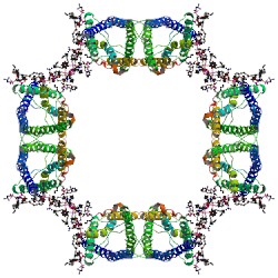
electron crystallographic structure of lens Aquaporin-0 (AQP0) (lens MIP) at 1.9A resolution, in a closed pore state
2B6O
Primary Citation

Lipid-protein interactions in double-layered two-dimensional AQP0 crystals.

Gonen, T.,  Cheng, Y.,  Sliz, P.,  Hiroaki, Y.,  Fujiyoshi, Y.,  Harrison, S.C.,  Walz, T.

Journal: (2005) Nature 438: 633-638

PubMed: 16319884  
PubMedCentral: PMC1350984  
DOI: 10.1038/nature04321  
Search Related Articles in PubMed  
PubMed Abstract:
Lens-specific aquaporin-0 (AQP0) functions as a specific water pore and forms the thin junctions between fibre cells. Here we describe a 1.9 A resolution structure of junctional AQP0, determined by electron crystallography of double-layered two-dimensional crystals. Comparison of junctional and... [ Read More & Search PubMed Abstracts ]
 
  •   Molecular Description Hide
    Classification: Membrane Protein
    Structure Weight: 34386.56
    Molecule: Lens fiber major intrinsic protein
    Polymer: 1 Type: protein Length: 263
    Chains: A
    Organism Ovis aries
    Gene Names MIP AQP0
    UniProtKB:   Protein Feature View | Search PDB | Q6J8I9  
    Q6J8I9Molec. ProcessingLens fiber major intrinsic proteinMotifCyHelicalEHelicaliHeCHelicalExtrHelicalCytHeliiHeEHelicalCytoplasmicUP SitesUniProtKBSCOP domainsAquaporin-like PDB SitesSecstruc2B6O.APDBTooltip
     
  •   Source Hide
    Polymer: 1
    Scientific Name: Ovis aries   Taxonomy   Common Name: Sheep  
     
  •   Related PDB Entries Hide
    Identifier Details
    1SOR  3A resolution structure of lens aquaporin 0 (AQP0; MIP) in a closed pore state determined by electron crystallography. 
    1YMG  2.2A resolution structure of lens aquaporin0 (AQP0; MIP) in an open pore state determined by Xray crystallography. 
    2B6P  2.5A resolution structure of lens aquaporin0 (AQP0; MIP) in an open pore state determined by Xray crystallography. 
     
  •   Ligand Chemical Component Hide
    Identifier Formula Name Interactions
    MC3
    Search 
    Download 
    MC3 C36 H72 N O8 P
    1,2-DIMYRISTOYL-RAC-GLYCERO-3-PHOSPHOCHOLINE
     
  •   External Domain Annotations Hide
     
  •   Structural Biology Knowledgebase Data Hide

    Information from the Structural Biology Knowledgebase  


Data in orange boxes are gathered from external resources (when available).
 
  Biological Assembly 1       
Biological assembly 1 assigned by authors
Downloadable viewers:


  •   MyPDB Personal Annotations Hide

    To save personal annotations, please login to your MyPDB account.

     
  •   Deposition Summary Hide
    Authors:   Gonen, T.,  Cheng, Y.,  Sliz, P.,  Hiroaki, Y.,  Fujiyoshi, Y.,  Harrison, S.C.,  Walz, T.

    Deposition:   2005-10-03
    Release:   2005-12-06
    Last Modified (REVDAT):   2011-07-13
     
  •   Revision History    Hide
    Mouse over text for details
    2011-07-13
    Version format compliance
    2011-07-13
    Biological assembly
     
  •   Experimental Details Hide
    Method:   ELECTRON CRYSTALLOGRAPHY
    Exp. Data:
      Structure Factors
    EDS  


    Space Group: P 4 2 2
    Unit Cell:
      Length [Å] Angles [°]
    a = 65.50 α = 90.00 
    b = 65.50 β = 90.00 
    c = 160.00 γ = 90.00