POP-OUT | CLOSE
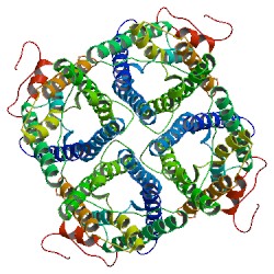
X-ray structure of lens Aquaporin-0 (AQP0) (lens MIP) in an open pore state
2B6P
Primary Citation

Lipid-protein interactions in double-layered two-dimensional AQP0 crystals.

Gonen, T.,  Cheng, Y.,  Sliz, P.,  Hiroaki, Y.,  Fujiyoshi, Y.,  Harrison, S.C.,  Walz, T.

Journal: (2005) Nature 438: 633-638

PubMed: 16319884  
PubMedCentral: PMC1350984  
DOI: 10.1038/nature04321  
Search Related Articles in PubMed  
PubMed Abstract:
Lens-specific aquaporin-0 (AQP0) functions as a specific water pore and forms the thin junctions between fibre cells. Here we describe a 1.9 A resolution structure of junctional AQP0, determined by electron crystallography of double-layered two-dimensional crystals. Comparison of junctional and... [ Read More & Search PubMed Abstracts ]
 
Original Entry Used for Re-refinement
1YMG 2.2A resolution structure of lens aquaporin0 (AQP0; MIP) in an open pore state determined by Xray crystallography. THIS ENTRY 2B6P REFLECTS AN ALTERNATIVE MODELING OF X-RAY DATA R1YMGSF 

The Channel Architecture Of Aquaporin O At 2.2 Angstrom Resolution
Harries, W.E.C., Akhavan, D., Miercke, L.J.W., Khademi, S., Stroud, R.M.
(2004) Proc.Natl.Acad.Sci.USA 101: 14045
PubMed
 
  •   Molecular Description Hide
    Classification: Membrane Protein
    Structure Weight: 28245.10
    Molecule: Lens fiber major intrinsic protein
    Polymer: 1 Type: protein Length: 263
    Chains: A
    Organism Bos taurus
    Gene Name MIP
    UniProtKB:   Protein Feature View | Search PDB | P06624  
    P06624Molec. ProcessingLens fiber major intrinsic proteinMotifCyHelicalEHelicaliHeCHelicalExtrHelicalCytHeliiHeEHelicalCytoplasmicUP SitesUniProtKBSCOP domainsAquaporin-like Secstruc2B6P.APDBTooltip
     
  •   Source Hide
    Polymer: 1
    Scientific Name: Bos taurus   Taxonomy   Common Name: Cattle  
     
  •   Related PDB Entries Hide
    Identifier Details
    1SOR  3A resolution structure of lens aquaporin 0 (AQP0; MIP) in a closed pore state determined by electron crystallography. 
    2B6O  1.9A resolution structure of lens aquaporin 0 (AQP0; MIP) in a closed pore state determined by electron crystallography. 
     
  •   External Domain Annotations Hide
     
  •   Structural Biology Knowledgebase Data Hide

    Information from the Structural Biology Knowledgebase  


Data in orange boxes are gathered from external resources (when available).
 
  Biological Assembly       
Biological assembly 1 assigned by authors and generated by PISA (software)
Downloadable viewers:


  •   MyPDB Personal Annotations Hide

    To save personal annotations, please login to your MyPDB account.

     
  •   Deposition Summary Hide
    Authors:   Gonen, T.,  Cheng, Y.,  Sliz, P.,  Hiroaki, Y.,  Fujiyoshi, Y.,  Harrison, S.C.,  Walz, T.

    Deposition:   2005-10-03
    Release:   2005-12-06
    Last Modified (REVDAT):   2011-10-26
     
  •   Revision History    Hide
    Mouse over text for details
    2011-10-26
    Other
    2011-07-13
    Version format compliance
     
  •   Experimental Details Hide
    Method:   X-RAY DIFFRACTION
    Exp. Data:
      Structure Factors
    EDS  
    Resolution[Å]:   2.40
    R-Value: 0.242 (work)
    R-Free: 0.292
    Space Group: P 4 21 2
    Unit Cell:
      Length [Å] Angles [°]
    a = 109.53 α = 90.00 
    b = 109.53 β = 90.00 
    c = 52.82 γ = 90.00