Advanced Biochemistry Fall Term, 1996
 Green Fluorescent
Protein Links for Problem Set #12
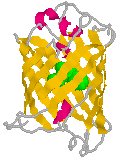
GFP is colored by "Structure".
The chromophore is shown in green.
(From the 1ema.pdb coordinate file.)
GFP Links
The following sites provide images and information on green fluorescent protein:
- The Molecular Structure of Green Fluorescent Protein. An on-line version of Yang et al. (1996) Nature Biotechnology14 1246-1251 with color images. This page takes a while to load, but the color figures are worth the wait.
Alternatively, to view the figures without captions, click on the following:
Figure 1 (50K); Figure 2 (36K); Figure 3 (27K);
Figure 4 (86K); Figure 5 (36K); Figure 6 (32K). - An active Chime image of GFP. Coordinates and description based on Ormo et al. (1996) Science273 1392-1395. (Requires the Chime plug-in and Netscape 3.0.)
Buttons corresponding to student-submitted RasMol scripts of GFP have been added to the page.
- Green Fluorescent Protein. A product description of cloning vectors from Clontech Inc. with references and some photos of green cells.
Another page at the same company describes an "Optimized GFP" that produces 18-fold more light than the wild type GFP.
Another mutant with the S65T and F64L substitutions was described recently by Yang et al. (1996) Nucleic Acids Res. 24 4592. Its fluorescence is about 35-fold higher than wild type due to a comparable increase in excitation coefficient at 488 nm. - A sampling (Medline Abstracts) of references to various uses for Green Fluorescent Protein from the Jackson Laboratories.
- Aequorea victoria, without whom this problem set would not have been possible!
 Return to ABC96 Home Page
Last modified December 9, 1996.
Green Fluorescent
Protein Links for Problem Set #12
Return to ABC96 Home Page