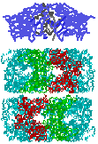
 GroE Structures and Architecture
GroE Structures and Architecture
GroE Structures and Architecture
A GroES dome caps the GroEL
"double-donut" to become a
protein folding machine
powered by ATP hydrolysis.
The pictures on this page are intended to complement the overview article by Lorimer & Todd, "GroE structures galore" Nature Struct. Biol.3, 116-121 (1996). The material is organized in three sections:
In addition, the following PDB files (also available in the Adv. Biochem. volume of the BioServer) can be used to view GroEL in RasMac. To view these structures directly from this page, click on the linked file name.
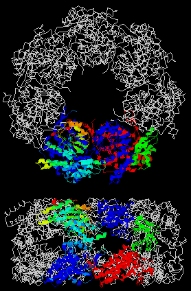 |
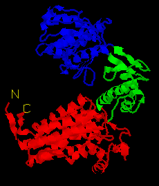
Image 1: Top and side views of the GroEL heptamer. (from 1oel.pdb)
1. The colored subunit on the left shows the crystallographic B-factor increasing from
blue shades (B=5) to red (B=150) using RasMac "Temperature" coloring.
2. The colored subunit on the right shows the domain organization of GroEL:
Note the correspondence between the B-factor coloring on the left and the domain organization shown on the right. Click on domain organization of GroEL for a larger picture of this subunit. It is a cartoon display made from 1oelSubA.pdb; the view is similar to that shown in Fig. 4 of Lorimer & Todd (1996).
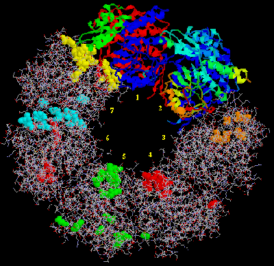 |
Image 2: Top view of the GroEL ring. (from 1oel.pdb)
Starting at the top of the picture, subunits 1 & 2 are shown in the "Domain" and "Temperature" coloring explained in Image 1. The remaining five subunits, displayed as wireframe, show the location of residues involved in various binding interactions.The amino acid residues are displayed as spacefill and are colored as follows:
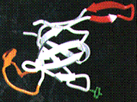 |
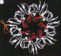
GroES subunit structure.
Fig. 1 from Lorimer & Todd (1996). The mobile loop is shown in orange; the ![]() -barrel core is shown
in white; and the
-barrel core is shown
in white; and the ![]() -hairpin
roof appendage is shown in red. See their caption for additional details.
-hairpin
roof appendage is shown in red. See their caption for additional details.
 |
Top View of the GroES dome architecture.
Fig. 2 from Lorimer & Todd (1996). Same coloring scheme described above. See their caption for additional details.
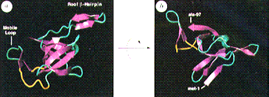 |
Orthogonal views of the GroES subunit structure.
Fig. 2 from Hunt et al. (1996); the mobile loop is resolved in only one of the
seven subunits. The linking segments between that ![]() -hairpin and the
-hairpin and the ![]() -barrel core structure are shown in yellow. The
other linking segments between
-barrel core structure are shown in yellow. The
other linking segments between ![]() -strands are shown in cyan. See their caption for additional
details.
-strands are shown in cyan. See their caption for additional
details.
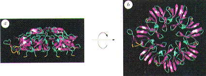 |
Side (a) and top (b) views of the GroES dome.
Fig. 3 from Hunt et al. (1996); same coloring scheme described above.
![]() Return to ABC97 Home Page
Return to ABC97 Home Page
Last modified November 11, 1996.