POP-OUT | CLOSE
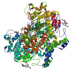
X-ray crystal structure of docosahexaenoic acid bound to the cyclooxygenase channel of cyclooxygenase-2
3HS7
Primary Citation

Structural basis of fatty acid substrate binding to cyclooxygenase-2.

Vecchio, A.J.,  Simmons, D.M.,  Malkowski, M.G.

Journal: (2010) J.Biol.Chem. 285: 22152-22163

PubMed: 20463020  
PubMedCentral: PMC2903402  
DOI: 10.1074/jbc.M110.119867  
Search Related Articles in PubMed  
PubMed Abstract:
The cyclooxygenases (COX-1 and COX-2) are membrane-associated heme-containing homodimers that generate prostaglandin H(2) from arachidonic acid (AA). Although AA is the preferred substrate, other fatty acids are oxygenated by these enzymes with varying efficiencies. We determined the crystal structures of... [ Read More & Search PubMed Abstracts ]
 
 
Data in orange boxes are gathered from external resources (when available).
 
  Biological Assembly       
Symmetry: C2 view
Stoichiometry: Homo 2-mer - A2
Biological assembly 1 assigned by authors and generated by PISA (software)
Downloadable viewers:


  •   MyPDB Personal Annotations Hide

    To save personal annotations, please login to your MyPDB account.

     
  •   Deposition Summary Hide
    Authors:   Vecchio, A.J.,  Simmons, D.M.,  Malkowski, M.G.

    Deposition:   2009-06-10
    Release:   2010-05-12
    Last Modified (REVDAT):   2010-08-04
     
  •   Revision History    Hide
    Mouse over text for details
    2011-07-13
    Version format compliance
     
  •   Experimental Details Hide
    Method:   X-RAY DIFFRACTION
    Exp. Data:
      Structure Factors
    EDS  
    Resolution[Å]:   2.65
    R-Value: 0.184 (obs.)
    R-Free: 0.234
    Space Group: I 2 2 2
    Unit Cell:
      Length [Å] Angles [°]
    a = 119.29 α = 90.00 
    b = 131.71 β = 90.00 
    c = 179.24 γ = 90.00