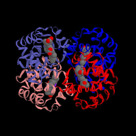
Ribbon Structure of Deoxy Fetal Hemoglobin (HbF)
Overview of Fetal Hemoglobin, HbF
Ribbon Structure of Deoxy Fetal Hemoglobin (HbF)
Fetal hemoglobin, HbF, is the predominant form of hemoglobin expressed in the developing fetus, appearing a few weeks post-conception and persisting for a few months post-birth. While having the same a-chain composition as adult hemoglobin, HbA, HbF is composed of two additional polypeptide chains, refered to as gamma- (g-) subunits, that are highly homologous to the b-chains of HbA. Thus, HbF is an a2g2 tetramer.
As a consequence of the structural differences between these two isoforms of hemoglobin, HbF exhibits a higher average affinity for oxygen than HbA. This is demonstrated by the fact that the O2 saturation curve for HbF is shifted to the left relative to the curve for HbA when tested under identical experimental conditions. This shift is physiologically meaningful because O2 must move from the maternal circulation to the fetal circulation and the higher average of affinity of HbF insures that O2 will ultimately flow in the direction of the fetal circulation.
The primary structural alterations in HbF as compared to HbA are found in or near the 2,3-BPG binding site. The net effect of these changes are that 2,3-BPG binds less tightly to deoxyHbF as compared to deoxyHbA. Thus, 2,3-BPG does not stabilize the deoxyHbF as effectively as it stabilizes deoxyHbA, thus accounting for the leftward shift of the O2 saturation curve of HbF compared to HbA when tested with the same concentration of 2,3-BPG present.
The structural locations of key differences between HbA and and HbF are summarized below.
| HbF g-chain (charge) | HbA b-chain (charge) | Net charge difference in or near HbF BPG pocket | |
| N-terminal Gly1-g acetylated (0) | N-terminal, a-NH3+of Val1-b (+1) | -2 | |
| Phe3-g (0) | Leu3-b (0) | 0 (Phe3-g pushes His2-g into BPG pocket changing the imiazole interactions with BPG) | |
| Asp80-g(-1) | Asn80-b (0) | -2 | |
| Ser143 (0) | His143-b (+1) | -2 | |
| Arg144 (+1) | Lys144 (+1) | 0 | |
| Net difference in charges on HbFg2 compared to HbAb2 | -6 | ||
Presumably BPG binding affinity at the g-ginterface of deoxyHbF is destablized by:
Structural images highlighting these changes are found
Fetal Hemoglobin HbF  Compared to HbA
| Go to top | |
| | Return to Biochemistry 108A Home Page |
| Press the browser's "Back" key to return to previous page. | |
| | |
© Duane W.
Sears
Revised: July 27, 1998