| Sarcomere; the functional contractile unit
|
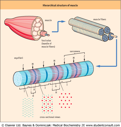
| Figure 19.1 Hierarchical structure of muscle. Hierarchical structure of skeletal muscle, showing an exploding view of fasciculi, myofibers, myofibrils and myofilament proteins. The location of the I-band (thin, actin filaments extending from a Z-line), and the A-band (thick, myosin filaments, extending from the M-line), with darker staining regions of the A-band corresponding to the region of overlap of actin and myosin filaments. |
| page 262 |  | | page 263 |
|
Table 19-1.
The structural elements of skeletal muscle arranged in descending order of size. |
| Body_ID: None |
| Elements of skeletal muscle structure |
| Body_ID: T019001.50 |
| Microscopic unit | Fasciculus: bundle of muscle cells |
| Body_ID: T019001.100 |
| Cellular unit | myofiber cell: long, multinucleated cell |
| Body_ID: T019001.150 |
| Subcellular unit | myofibril: composed of myofilament proteins |
| Body_ID: T019001.200 |
| Functional unit | sarcomere: contractile unit, repeating unit of the myofibril |
| Body_ID: T019001.250 |
| Myofilament components | proteins: primarily actin and myosin |
| Body_ID: T019001.300 |
| Unlike other cell types, the unique characteristic of muscle structure is that the cytoplasm is packed full of contractile protein. The arrangement of this contractile protein into sarcomere units gives muscle a striated appearance and has given rise to the classifications of striated (skeletal, cardiac) or non-striated muscle (smooth). Skeletal muscle's hierarchical structure (Fig. 19.1) consists of bundles (fasciculi) of elongated, multinucleated fiber cells (myofibers). The myofiber cells contain bundles of myofibrils, which are, in turn, composed of myofilament proteins that form the sarcomere (Table 19.1). Electron microscopic analysis reveals a repeating pattern of light- and dark-staining regions in the myofibril, which have been defined as specific, named bands (Fig. 19.2).
The light- and dark-staining regions are known as the I (isotropic)- and A (anisotropic)-bands, respectively. At the center of the I-band is a discrete, darker staining Z-line, while the center of the A-band has a lighter staining H-zone with a central M-line. The sarcomere, centered on the M-line, extends from one Z-line to the next. Smooth muscle lacks a defined Z-line in its sarcomere structure.
|
|