| N-linked oligosaccharides
|
| The assembly of the N-linked oligosaccharide chains begins in the endoplasmic reticulum
|
| page 366 |  | | page 367 |
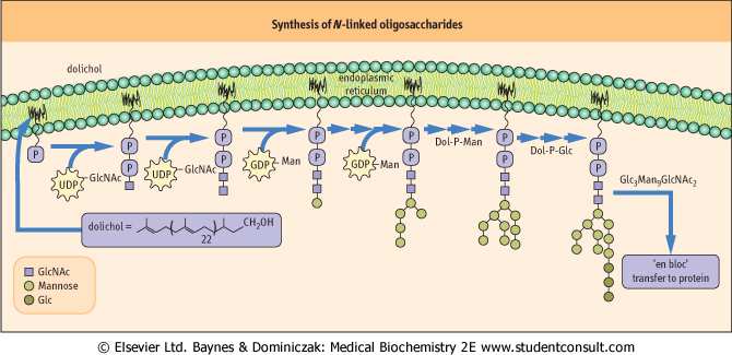
| Figure 25.10 Synthesis of N-linked oligosaccharides in the endoplasmic reticulum. GlcNAc, acetylglucosamine; Dol, dolichol; Man, mannose. |
| INHIBITORS OF GLYCOSYL TRANSFERASES |
| Several inhibitors of the biosynthesis of N-linked oligosaccharides have become valuable reagents for understanding the role of specific carbohydrate structures in glycoprotein function. Tunicamycin is a glycoside antibiotic that inhibits the first step in synthesis of N-linked oligosaccharides - formation of Dol-PP-GlcNAc (see Fig. 25.10). It has varied effects on the synthesis and function of glycoproteins. Some proteins are insoluble without their carbohydrate, and aggregate in the cell. Others do not fold correctly, or may fail to function in recognition reactions. Tunicamycin is quite toxic to animals, as animal cells need N-linked oligosaccharides for essential functions. |
The pathway of assembly of N-linked oligosaccharides involves the participation of a lipid carrier to form an activated oligosaccharide, which is then transferred en bloc to specific asparagine residues on the protein (Fig. 25.10). The lipid carrier is a long-chain polyisoprenol of about 120 carbon atoms, with a phosphate group esterified to the terminal isoprene unit. This molecule, called dolichol phosphate (Dol-P), is an integral lipid of the endoplasmic reticulum. The individual sugars - GlcNAc, mannose and glucose - are added to Dol-P, one at a time, to form Glc3Man9GlcNAc2-PP-Dol, which then donates the oligosaccharide to protein. For
N-linked oligosaccharides, glycosylation is co-translational, which means that it occurs while the peptide chain is still being synthesized on the membrane-bound ribosome (see Chapter 32). - are added to Dol-P, one at a time, to form Glc3Man9GlcNAc2-PP-Dol, which then donates the oligosaccharide to protein. For
N-linked oligosaccharides, glycosylation is co-translational, which means that it occurs while the peptide chain is still being synthesized on the membrane-bound ribosome (see Chapter 32).
|
| The first GlcNAc residue is added with its phosphate residue, so that the core structure is dolichyl pyrophosphate-GlcNAc (Dol-PP-GlcNAc), a hydrophobic, membrane-bound analogue of a sugar nucleotide. The remaining GlcNAc and five mannose units are transferred from their sugar nucleotides (UDP-GlcNAc and GDP-Man), whereas the next four mannose and three glucose residues are donated from lipid precursors (Dol-P-mannose and Dol-P-glucose). Each of the sugars that are added to the Dol-PP carrier is transferred by a specific enzyme; these enzymes are members of the class of enzymes known as glycosyl transferases. The completed oligosaccharide is finally transferred from its Dol-PP derivative to an asparagine residue in an Asn-X-Ser(Thr) sequence in a protein.
|
| The function of the glucoses on the initial oligosaccharide is to expedite oligosaccharide transfer from lipid to protein. Oligosaccharyl transferase, the transferring enzyme, has a preference for oligosaccharides that contain three glucose units. In addition, as indicated below, the glucose residues are important for expediting the folding of the protein.
|
| Intermediate processing continues in the endoplasmic reticulum
|
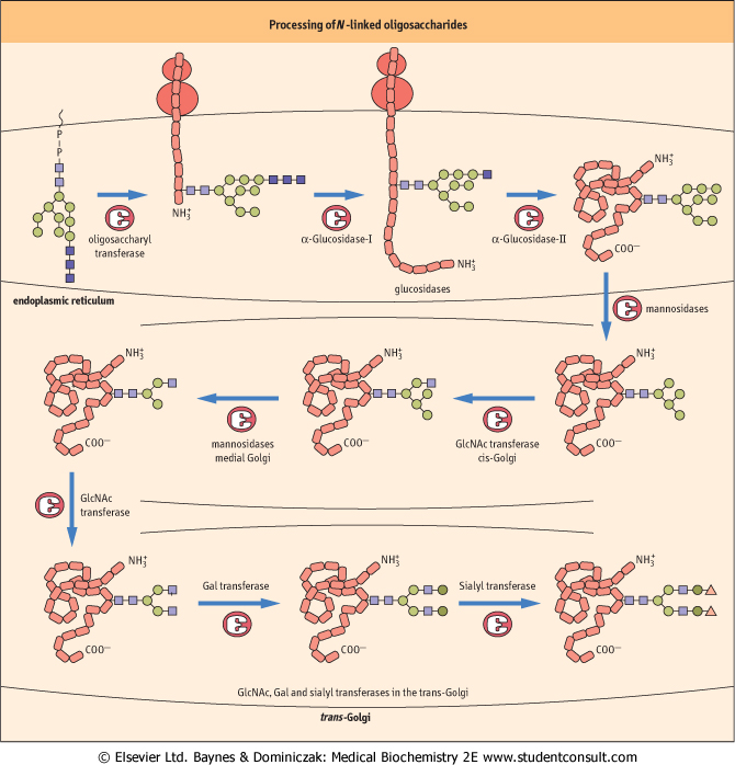
| Once the oligosaccharide chain has been transferred to the protein, various glycosidases (enzymes that remove specific sugars, including the glucosidases that are specific for glucose) act on the protein-bound oligosaccharide. In a series of processing or 'pruning' reactions, the three glucose residues are removed in the endoplasmic reticulum, and up to six mannose residues in the Golgi apparatus. These trimming reactions give rise to a core structure of two GlcNAc and three mannose residues, and this core oligosaccharide is elongated to form complex oligosaccharides. The elongation reactions involve the addition of one or more of the sugars, GlcNAc, galactose, sialic acid, and l-fucose. The reactions involved in the modification of the oligosaccharide chains are outlined in Figure 25.11.
|
| page 367 | | | page 368 |
| Figure 25.11 Processing of N-linked oligosaccharides from high-mannose to complex forms. Glycoproteins are transported between the endoplasmic reticulum and Golgi compartments in vesicles. GlcNAc, acetylglucosamine. |
| page 368 | | | page 369 |
| Final modifications to the glycoprotein take place in the Golgi apparatus
|
| When the glucose residues have been removed and the protein has folded into its correct conformation, the glycoprotein with one or more Man9GlcNAc2 oligosaccharides is transported from the endoplasmic reticulum to the Golgi apparatus, where other modification reactions can occur. Usually, in the cis and medial regions of the Golgi, six of the mannose residues are removed by several different α-mannosidases. The exact role of each of the mannosidases is not known, but they produce glycoproteins with oligosaccharide chains having variable numbers of mannose residues. If the oligosaccharide is trimmed to its core structure, it may then be converted to a complex structure by glycosyl transferases in the trans-Golgi apparatus (Fig. 25.11), which add GlcNAc, Gal, sialic acid and fucose residues.
|
|