| Factors involved in the elongation stage of protein synthesis are targets of some antibiotics
|
| A young man you were treating for a sinus infection returns to your clinic after 1 week, still complaining of sinus headaches and stuffiness. He explains that he began to feel better about 3 days after starting to take the antibiotic tetracycline, which you had prescribed. You inquire as to whether he continued to take the full dose of the drug, even after he began to feel better. He reluctantly admits that, as soon as he felt better, he stopped taking the drug. How do you explain to your patient that it is important that he takes the drug for as long as you prescribed it, even if he feels better after only a few days? |
| Comment. As a physician, you know that tetracycline inhibits the protein synthetic machinery of the bacterial cell by binding to the A site of the ribosome (Table 32.4). You also know that, if the drug is removed, protein synthesis can resume. If the drug is not taken for the entire period recommended, bacteria will begin to grow again, leading to the resurgence of the infection. Further, those bacteria that begin to grow after early termination of treatment are likely to be the most resistant to the drug, either because of selection of more resistant strains, or because of mutation to more resistant strains. The secondary infection is therefore likely to be more difficult to control. |
| PROTEIN SYNTHESIS: PEPTIDYL TRANSFERASE |
| Peptidyl transferase is not your typical enzyme. It is a ribozyme |
| Peptidyl transferase is the enzyme responsible for peptide bond formation during protein synthesis. This enzyme catalyzes the reaction between the amino group of the aminoacyl-tRNA, forming a peptide bond from an ester bond. The enzyme activity is located in the ribosome, but none of the ribosomal proteins has the capacity to catalyze this reaction. In fact, when ribosomes are stripped of all of their associated proteins, the rRNA appears to remain capable of converting an ester bond to a peptide bond. These observations have led investigators to hypothesize that peptidyl transferase activity is contained in the rRNA, rather than in the proteins that associate with ribosomes. |
| page 452 |  | | page 453 |
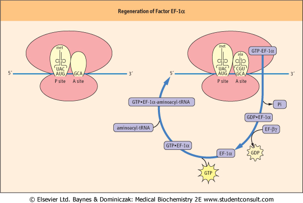
| Figure 32.5 Recycling of Factor EF-1α. A charged tRNA molecule is brought to the A site of the initiation complex to begin the process of elongation. Each successive amino acid addition requires that the correctly charged tRNA molecule be brought to the A site of the ribosome. ala, alanine; Pi, inorganic phosphate. |
| After initiation is complete, the process of translating the information in mRNA into a functional protein starts. Elongation begins with the binding of a charged tRNA to the
A site of the ribosome. In eukaryotic cells, the charged tRNA molecule is brought to the ribosome by the action of an elongation factor called EF-1α (Fig. 32.5). For EF-1α to be active, it must have a GTP molecule associated with it. If the charged tRNA is the correct one - i.e., one in which the anticodon of
the tRNA forms base pairs with the codon on the mRNA - GTP is hydrolyzed and EF-1α is released. For EF-1α factor to bring another charged molecule to the ribosome, it must be regenerated by an elongation factor called EF-βγ, which will promote the association of EF-1α with GTP so that it may bind to another charged tRNA molecule (Fig. 32.5). Once the correct charged tRNA molecule has been delivered to the A site of the ribosome, peptidyl transferase catalyzes the formation of a peptide bond between the amino acid in the A site and the amino acid at the end of the growing peptide chain in the P site. The tRNA-peptide chain is now transiently bound to the A site. The ribosome is then moved one codon down the mRNA by a factor known as EF-2 and the nascent peptide chain at the A site moves to the P site. The whole process recycles for addition of the next amino acid (Fig. 32.6). This complex process is identical in prokaryotic cells, but the factors are different; this explains the utility of antibiotics that preferentially inhibit protein synthesis in bacteria (Table 32.4).
|
|
Table 32-4.
Antibiotics and their targets. |
| Body_ID: None |
| Antibiotics and their targets |
| Body_ID: T032004.50 |
| Antibiotic | Target |
| Body_ID: T032004.100 |
| tetracycline | bacterial ribosome-A site |
| Body_ID: T032004.150 |
| streptomycin | bacterial 30 S ribosome subunit |
| Body_ID: T032004.200 |
erythromycin | bacterial 50 S ribosome subunit |
| Body_ID: T032004.250 |
| chloramphenicol | bacterial ribosome-peptidyl transferase |
| Body_ID: T032004.300 |
| cycloheximide | eukaryotic 80 S ribosome |
| Body_ID: T032004.350 |
| ricin | eukaryotic 60 S ribosome subunit |
| Body_ID: T032004.400 |
|
| Body_ID: T032004.450 |
Cycloheximide, and ricin in particular, are potent poisons to humans.
|
|