| The term blood-brain 'barrier' is a slight misnomer, in that the barrier is not absolute, but relative: its permeability depends on the size of the molecule in question
|
| Initially, experiments based upon use of a dye (Evans' Blue) bound to albumin showed that, over a period of hours, an animal progressively turned blue in all tissues, with the notable exception of the brain, which remained white. It subsequently became clear that 1 molecule in 200 of serum albumin passed normally into the CSF, which is analogous to lymph. It also became obvious that, for any given protein, the ratio of its concentrations in CSF and serum was a linear function of the molecular radius of the molecules in solution.
|
| page 559 |  | | page 560 |
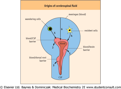
| There are a total of six sources of the CSF which contain different 'barriers'. Under normal and pathologic conditions, proteins pass from these cellular or tissue sources into the
CSF, and their degrees of filtration or rates of local synthesis, or both, vary.
|
The total quantity of the CSF therefore constitutes the algebraic summation of these six sources (Fig. 39.1):
- the blood-brain barrier (the parenchymal capillaries) gives rise to about one-third of the volume of CSF, and has been termed the interstitial fluid source;
- the blood-CSF barrier provides the bulk of CSF (almost all of the remaining two-thirds), termed choroidal fluid, as it is principally provided by the choroid plexi (capillary tufts) situated in the lateral ventricles and, to a lesser degree, the plexi situated in the third and fourth ventricles;
- the dorsal root ganglia contain capillaries that have a much greater degree of permeability. In the animal experiments with the Evans' blue referred to above, although the brain was otherwise white, the dorsal roots took up the blue color reflecting their greater permeability to albumin;
- the brain parenchyma of the CNS produces a number of brain-specific proteins. These include prostaglandin synthase (formerly called β-trace protein), which shows an 11-fold increase from the choroid plexus to the lumbar sac, and transthyretin (a protein formerly called prealbumin which is produced locally by the choroid plexi), for which the reverse is found: it has a much greater relative concentration in ventricular than in lumbar CSF;
- CSF circulating cells, mainly lymphocytes within the CNS, synthesize local antibodies: however, in the CNS there is strong presence of immune suppressor cells. Because of this, in brain infections such as meningitis, steroids are given in addition to antibiotics, to suppress the potentially devastating effects, within this confined space, of inflammation associated with the intrathecal immune response;
- the meninges represent a sixth source of CSF under pathologic conditions; they can give rise to dramatic increase in the concentrations of CSF proteins.
|
| Figure 39.1 The six main sources of cerebrospinal fluid (CSF). The involved processes comprise passage across barriers (from the blood, i.e. 1,2,3) and direct sources of local production (CNS cells, i.e. 4,5,6). |
|