POP-OUT | CLOSE
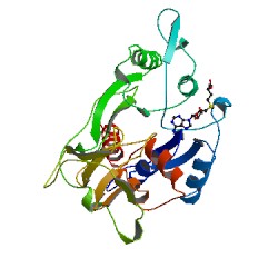
CRYSTAL STRUCTURE OF THE HHAI DNA METHYLTRANSFERASE COMPLEXED WITH S-ADENOSYL-L-METHIONINE
1HMY
Primary Citation

Crystal structure of the HhaI DNA methyltransferase complexed with S-adenosyl-L-methionine.

Cheng, X.,  Kumar, S.,  Posfai, J.,  Pflugrath, J.W.,  Roberts, R.J.

Journal: (1993) Cell(Cambridge,Mass.) 74: 299-307

PubMed: 8343957  
Search Related Articles in PubMed  
PubMed Abstract:
The first three-dimensional structure of a DNA methyltransferase is presented. The crystal structure of the DNA (cytosine-5)-methyltransferase, M.HhaI (recognition sequence: GCGC), complexed with S-adenosyl-L-methionine has been determined and refined at 2.5 A resolution. The core of the structure is dominated... [ Read More & Search PubMed Abstracts ]
 
  •   Molecular Description Hide
    Classification: Transferase(methyltransferase)
    Structure Weight: 37440.94
    Molecule: HaeIII METHYLTRANSFERASE
    Polymer: 1 Type: protein Length: 327
    Chains: A
    EC#: 2.1.1.37   
    Organism Haemophilus haemolyticus
    Gene Name hhaIM
    UniProtKB:   Protein Feature View | Search PDB | P05102  
    P05102Molec. ProcessingModification methylase HhaIE.C.2.1.1.37: DNA (cytosine-5-)-methyltransferaseUP SitesUniProtKBSCOP domainsS-adenosyl-L-methionine-dependent methyltransferases PDB SitesSecstruc1HMY.APDBTooltip
     
  •   Source Hide
    Polymer: 1
    Scientific Name: Haemophilus haemolyticus   Taxonomy    
     
  •   Ligand Chemical Component Hide
    Identifier Formula Name Interactions
    SAM
    Search 
    Download 
    SAM C15 H22 N6 O5 S
    S-ADENOSYLMETHIONINE
    SAM:1HMY
     
  •   External Domain Annotations Hide
     
  •   Structural Biology Knowledgebase Data Hide

    Information from the Structural Biology Knowledgebase  


Data in orange boxes are gathered from external resources (when available).
 
 
  •   MyPDB Personal Annotations Hide

    To save personal annotations, please login to your MyPDB account.

     
  •   Deposition Summary Hide
    Authors:   Cheng, X.

    Deposition:   1993-08-05
    Release:   1993-10-31
    Last Modified (REVDAT):   2012-02-01
     
  •   Revision History    Hide
    Mouse over text for details
    2012-02-01
    Entry title
    2011-07-13
    Version format compliance
     
  •   Experimental Details Hide
    Method:   X-RAY DIFFRACTION
    Exp. Data:
    N/A
    Resolution[Å]:   2.50
    R-Value: 0.200 (obs.)
    R-Free: n/a
    Space Group: P 21
    Unit Cell:
      Length [Å] Angles [°]
    a = 55.30 α = 90.00 
    b = 72.70 β = 102.50 
    c = 91.00 γ = 90.00