POP-OUT | CLOSE
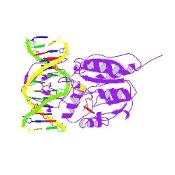
COVALENT TERNARY STRUCTURE OF HHAI METHYLTRANSFERASE, DNA AND S-ADENOSYL-L-HOMOCYSTEINE
DOI:10.2210/pdb1mht/pdb   NDB ID: PDEB08
1MHT
Primary Citation

HhaI methyltransferase flips its target base out of the DNA helix.

Klimasauskas, S.,  Kumar, S.,  Roberts, R.J.,  Cheng, X.

Journal: (1994) Cell(Cambridge,Mass.) 76: 357-369

PubMed: 8293469  
Search Related Articles in PubMed  
PubMed Abstract:
The crystal structure has been determined at 2.8 A resolution for a chemically-trapped covalent reaction intermediate between the HhaI DNA cytosine-5-methyltransferase, S-adenosyl-L-homocysteine, and a duplex 13-mer DNA oligonucleotide containing methylated 5-fluorocytosine at its target. The DNA is located in a... [ Read More & Search PubMed Abstracts ]
 
  •   Molecular Description Hide
    Classification: Transferase/dna
    Structure Weight: 45156.04
    Molecule: DNA (5'-D(P*GP*AP*TP*AP*GP*(C36)P*GP*CP*TP*AP*TP*C)-3')
    Polymer: 1 Type: dna Length: 12
    Chains: B
    Molecule: DNA (5'-D(*TP*GP*AP*TP*AP*GP*(C36)P*GP*CP*TP*AP*TP*C)-3')
    Polymer: 2 Type: dna Length: 13
    Chains: C
    Molecule: PROTEIN (HHAI METHYLTRANSFERASE)
    Polymer: 3 Type: protein Length: 327
    Chains: A
    EC#: 2.1.1.37   
    Organism Haemophilus haemolyticus
    Gene Name hhaIM
    UniProtKB:   Protein Feature View | Search PDB | P05102  
    P05102Molec. ProcessingModification methylase HhaIE.C.2.1.1.37: DNA (cytosine-5-)-methyltransferaseUP SitesUniProtKBSCOP domainsS-adenosyl-L-methionine-dependent methyltransferases PDB SitesSecstruc1MHT.APDBTooltip
     
  •   Source Hide
    Polymer: 1
    Scientific Name: Synthetic construct   Taxonomy    
    Polymer: 2
    Scientific Name: Synthetic construct   Taxonomy    
    Polymer: 3
    Scientific Name: Haemophilus haemolyticus   Taxonomy    
     
  •   Ligand Chemical Component Hide
    Identifier Formula Name Interactions
    SAH
    Search 
    Download 
    SAH C14 H20 N6 O5 S
    S-ADENOSYL-L-HOMOCYSTEINE
    SAH:1MHT
     
  •   Modified Residues Hide
    Identifier Formula Parent Type
    C36
    Search 
    C36 C10 H17 F N3 O8 P DC dnaLinking
     
  •   External Domain Annotations Hide
     
  •   Structural Biology Knowledgebase Data Hide

    Information from the Structural Biology Knowledgebase  

     
  • Nucleic Acid Database Hide
    View the NDB ID associated with this structure: PDEB08
     
 
Data in orange boxes are gathered from external resources (when available).
 
 
  •   MyPDB Personal Annotations Hide

    To save personal annotations, please login to your MyPDB account.

     
  •   Deposition Summary Hide
    Authors:   Cheng, X.

    Deposition:   1994-12-08
    Release:   1995-06-03
    Last Modified (REVDAT):   2009-02-24
     
  •   Revision History    Hide
    Mouse over text for details
    2011-07-13
    Version format compliance
     
  •   Experimental Details Hide
    Method:   X-RAY DIFFRACTION
    Exp. Data:
    N/A
    Resolution[Å]:   2.60
    R-Value: 0.174 (obs.)
    R-Free: n/a
    Space Group: H 3 2
    Unit Cell:
      Length [Å] Angles [°]
    a = 99.86 α = 90.00 
    b = 99.86 β = 90.00 
    c = 325.20 γ = 120.00