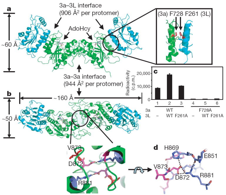
a, Side view of the tetramer Dnmt3L (blue)-Dnmt3a (green)-Dnmt3a (green)-Dnmt3L (blue), with the bound AdoHcy shown as a stick model. The inset shows the two pairs of phenylalanine residues (F728 and F768 of Dnmt3a, and F261 and F301 of Dnmt3L) in the centre of the Dnmt3a-Dnmt3L interface.
b, Top view showing the Dnmt3a-Dnmt3a interface with two pairs of salt bridges formed between R881 and D872 (enlarged).
c, Activities of Dnmt3a2 and its point mutant F728A in the presence and absence of Dnmt3L. Error bars represent s.d. calculated from two independent experiments.
d, A network of polar interactions between two Dnmt3a molecules (coloured blue and purple) involves
R881-D872, D872-H869,
H869-E851 and E851-R881. Back to the tutorial Methylase