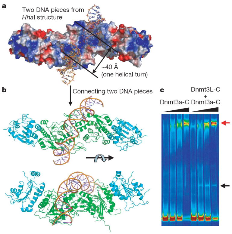
a, Surface representation of the Dnmt3a-Dnmt3L tetramer, with two short DNA molecules adopted by superimposition of the HhaI-DNA complex structure onto individual Dnmt3a-C.
b, Two views of the Dnmt3a-Dnmt3L tetramer with one contiguous curved DNA molecule, approximately 25 base pairs in length, covering two active sites with C.
c, Cooperative formation of large protein-DNA complexes (red arrow) after incubation of a 146-base-pair DNA with increasing concentrations of Dnmt3a-C or Dnmt3a-C-Dnmt3L-C complex. The lower band (black arrow) corresponds to one Dnmt3L-C bound to the DNA as a result of the presence of some free Dnmt3L-C in the mixture.
Back to the tutorial Methylase