POP-OUT | CLOSE
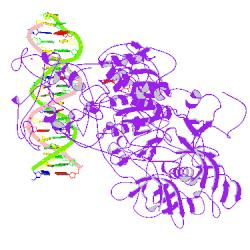
Crystal structure of mouse DNMT1(650-1602) in complex with DNA
DOI:10.2210/pdb3pt6/pdb   NDB ID: NA0856
3PT6
Primary Citation

Structure of DNMT1-DNA complex reveals a role for autoinhibition in maintenance DNA methylation.

Song, J.,  Rechkoblit, O.,  Bestor, T.H.,  Patel, D.J.

Journal: (2011) Science 331: 1036-1040

PubMed: 21163962  
DOI: 10.1126/science.1195380  
Search Related Articles in PubMed  
PubMed Abstract:
Maintenance of genomic methylation patterns is mediated primarily by DNA methyltransferase-1 (DNMT1). We have solved structures of mouse and human DNMT1 composed of CXXC, tandem bromo-adjacent homology (BAH1/2), and methyltransferase domains bound to DNA-containing unmethylated CpG sites. The CXXC specifically... [ Read More & Search PubMed Abstracts ]
 
  •   Molecular Description Hide
    Classification: Transferase/dna
    Structure Weight: 240683.06
    Molecule: DNA (cytosine-5)-methyltransferase 1
    Polymer: 1 Type: protein Length: 954
    Chains: A, B
    EC#: 2.1.1.37   
    Fragment: UNP residues 650-1602
    Organism Mus musculus
    Gene Names Dnmt1 Dnmt Met1 Uim
    UniProtKB:   Protein Feature View | Search PDB | P13864  
    P13864Molec. ProcessingDNA (cytosine-5)-methyltransferase 1MotifBAH 1BAH 2CInteraction wiAuCatalyticInteraInInteraction InteInteraction with theInterDNA repliNE.C.2.1.1.37: DNA (cytosine-5-)-methyltransferaseUP SitesUniProtKBPDB SitesSecstruc3PT6.B3PT6.APDBTooltip
    Molecule: DNA (5'-D(*TP*CP*CP*CP*GP*TP*GP*AP*GP*CP*CP*TP*CP*CP*GP*CP*AP*GP*G)-3')
    Polymer: 2 Type: dna Length: 19
    Chains: C, D
    Molecule: DNA (5'-D(*CP*CP*TP*GP*CP*GP*GP*AP*GP*GP*CP*TP*CP*AP*CP*GP*GP*GP*A)-3')
    Polymer: 3 Type: dna Length: 19
    Chains: I, J
     
  •   Source Hide
    Polymer: 1
    Scientific Name: Mus musculus   Taxonomy   Common Name: Mouse Expression System: Escherichia coli  
    Polymer: 2
    Scientific Name: Synthetic construct   Taxonomy    
    Polymer: 3
    Scientific Name: Synthetic construct   Taxonomy    
     
  •   Related PDB Entries Hide
    Identifier Details
    3PT9   
    3PTA   
     
  •   Ligand Chemical Component Hide
    Identifier Formula Name Interactions
    SAH
    Search 
    Download 
    SAH C14 H20 N6 O5 S
    S-ADENOSYL-L-HOMOCYSTEINE
    SAH:3PT6
    ZN
    Search 
    Download 
    ZN Zn
    ZINC ION
     
  •   External Ligand Annotations Hide
    Identifier   Binding Affinity (Sequence Identity %)
    SAH
    Search 
    Download 
    SAH
    IC50: 800 - 900 nM (85) - data from BindingDB  
    N/Ain BindingMoad
    N/Ain PDBbind
     
  •   External Domain Annotations Hide
     
  •   Structural Biology Knowledgebase Data Hide

    Information from the Structural Biology Knowledgebase  

     
  • Nucleic Acid Database Hide
    View the NDB ID associated with this structure: NA0856
     
 
Data in orange boxes are gathered from external resources (when available).
 
  Biological Assembly 1       
Biological assembly 1 assigned by authors and generated by PISA (software)
Downloadable viewers:


  •   MyPDB Personal Annotations Hide

    To save personal annotations, please login to your MyPDB account.

     
  •   Deposition Summary Hide
    Authors:   Song, J.,  Patel, D.J.

    Deposition:   2010-12-02
    Release:   2010-12-29
    Last Modified (REVDAT):   2011-03-30
     
  •   Revision History    Hide
    Mouse over text for details
    2011-07-13
    Version format compliance
     
  •   Experimental Details Hide
    Method:   X-RAY DIFFRACTION
    Exp. Data:
      Structure Factors
    EDS  
    Resolution[Å]:   3.00
    R-Value: 0.213 (obs.)
    R-Free: 0.266
    Space Group: P 21
    Unit Cell:
      Length [Å] Angles [°]
    a = 83.19 α = 90.00 
    b = 213.51 β = 99.64 
    c = 86.27 γ = 90.00