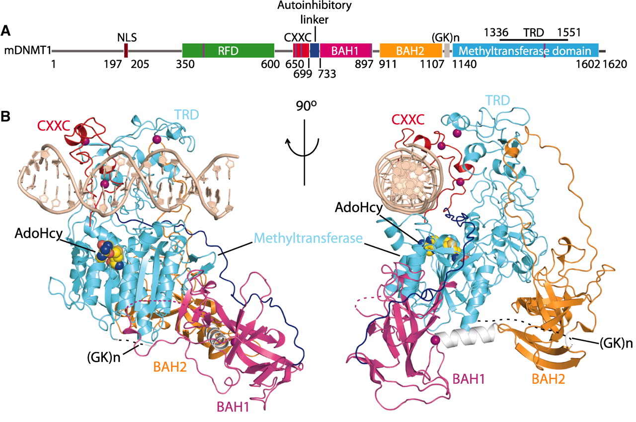
A) Color-coded domain architecture and numbering of mDNMT1 sequence. The thin vertical light blue bars indicate binding positions of zinc ions.
(B) Ribbon representation of the complex in two orthogonal views. The CXXC, BAH1, BAH2, and methyltransferase domain are colored in red, light purple, orange and light blue, and DNA and zinc ions are colored in light brown and dark purple, respectively; CXXC-BAH1 linker in dark blue, BAH1-BAH2 linker in silver, (GK)n-containing BAH2-methyltransferase linker in black, and bound AdoHcy as in space-filling representation.
Back to the tutorial Methylase