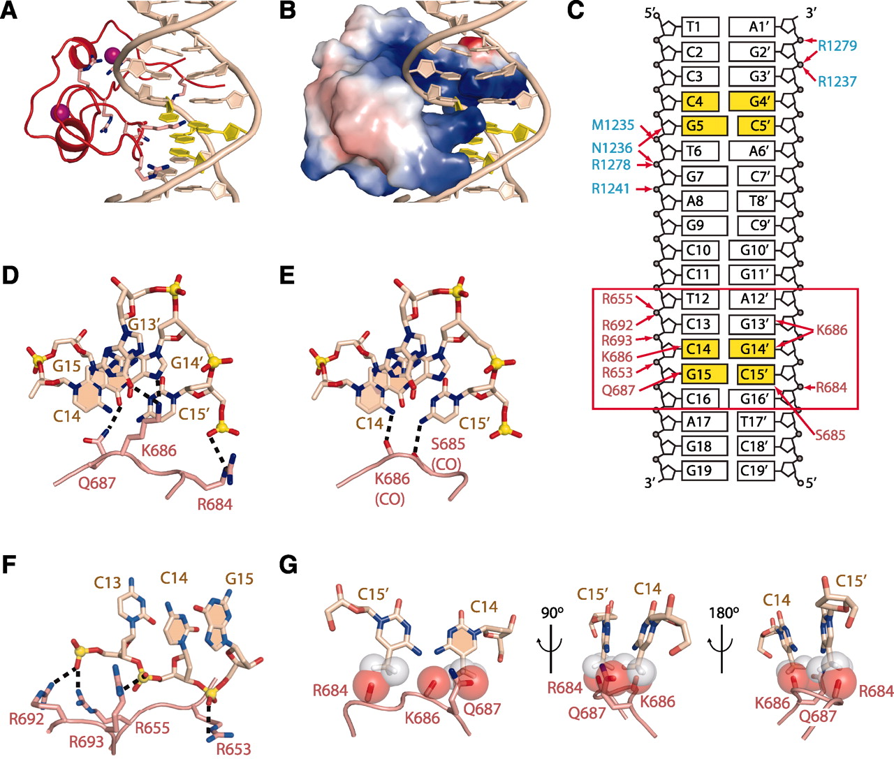
(A) Ribbon representation of the CXXCdomain bound to DNA. The CpG step is rendered in yellow.
(B) Surface electrostatic representation of the CXXCdomain bound to DNA.
(C) Schematic view of intermolecular interactions involving the CXXCdomain in the mDNMT1-DNA
19-nucleotide oligomer complex (boxed red rectangle), with intermolecular contacts shown by red arrows. The residue labels are colored according to their respective domains in Fig. 1.
(D) Hydrogen-bonding interactions between side chains from the CXXCdomain and the guanine base edges of the CpG step in the DNA major groove. The nitrogen, oxygen, and phosphorous atoms are shown in dark blue, red, and yellow, respectively. The bases of the CpG dinucleotide from one strand are shaded.
(E) Hydrogen-bonding interactions between backbone carbonyl oxygens from the CXXCdomain and cytosine amino groups of the CpG step in the DNA major groove.
(F) Hydrogen-bonding interactions between arginine side chains of the CXXCdomain and the phosphodiester backbone of the DNA along the DNA minor groove.
(G) Potential steric clashes between methylated cytosine modeled on either strand of the CpG step and the CXXCdomain of DNMT1 in the structure of the complex. Van der Waals radii are rendered in red for DNMT1 and gray for modeled methylated cytosine.
Back to the tutorial Methylase