| Immunoglobulins are proteins produced in response to foreign substances (antigens) (see also Chapter 36)
|
| The immune system may be conceptualized as two independent entities, served by separate lymphoid cells: thymically derived T lymphocytes oversee immunoregulation and cell-based immune function, and B lymphocytes that synthesize and secrete antibodies (immunoglobulins) (see Chapter 36). These antibodies are proteins, produced by the immune system, which have a defined specificity for a foreign particle (immunogen) that stimulated their synthesis. Not all foreign substances entering the body can elicit this response, however; those that do are called immunogens, whereas any agent that can be bound by an antibody is termed an antigen.
|
| The immunoglobulins are a uniquely diverse group of molecules, recognizing and reacting with a wide range of specific antigenic structures (epitopes) and giving rise to a series of effects that result in the eventual elimination of the presenting antigen. Some immunoglobulins have additional effector functions: for example, IgG is involved in complement activation.
|
| Structure of immunoglobulins
|
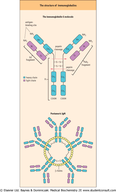
| Immunoglobulins share a common Y-shaped structure of two heavy and two light chains
|
| The basic immunoglobulin is a Y-shaped molecule containing two identical units termed heavy (H) chains, and two identical, but smaller, units termed light (L) chains. Several H chains exist, and the nature of the H chain determines the class of immunoglobulin: IgG, IgA, IgM, IgD, and IgE are characterized by γ, α, μ, δ, and ε heavy chains, respectively. L chains are of only two types κ and λ, and both types may be found in any one class of immunoglobulin, although obviously not within the same molecule. Each polypeptide chain within the immunoglobulin is characterized by a series of globular regions, which have considerable sequence homology and, in evolutionary terms, are probably derived from protogene duplication.
|
| The N-terminal domains of both H and L chains contain a region of variable amino acid sequence (the V region); together, these determine antigenic specificity. Both H and L chains are required for full antibody activity, as the physically apposed V regions in the L and H chains form a functional pocket into which the epitope fits; this is termed the antibody recognition (Fab2) region. The domain immediately adjacent to the V region is much less variable, in both H and L chains. The remainder of the H chain consists of a further constant region (Fc region) consisting of a hinge region and two additional domains. This constant region is responsible for immunoglobulin functions other than epitope recognition, such as complement activation (Chapter 36). This basic structure of immunoglobulins is depicted in Figure 3.6. When antigen binds to the immunoglobulin, conformational changes are transmitted through the hinge region of the antibody, to the Fc region, which is then said to have become activated.
|
| IgG is the most common immunoglobulin, protecting tissue spaces and freely crossing the placenta
|
| IgG 'with an overall molecular mass of 160 kDa' consists of the basic 2H2L immunoglobulin subunit joined by a variable number of disulfide bonds. The γ H chains have several antigenic and structural differences, allowing classification of IgG into a number of subclasses according to the type of H chain present; however, functional differences between the subclasses are minor.
|
| page 30 |  | | page 31 |
| Figure 3.6 The structure of immunoglobulins. Diagrammatic representation of the basic structure of a monomeric immunoglobulin and that of pentameric immunoglobulin (IgM). V, variable region; C, constant region; H, heavy chain; L, light chain; J chain, joining chain; Fab2, fragment generated by pepsin cleavage of the molecule; Fc, Fd, fragments generated by papain proteolysis (See also Chapter 36). |
| IgG circulates in high concentrations in the plasma, accounting for 75% of immunoglobulin present in adults, and has a half-life of 22 days. It is present in all extracelluar fluids, and appears to eliminate small, soluble antigenic proteins through aggregation and enhanced phagocytosis by the
reticuloendothelial system. From weeks 18-20 of pregnancy, IgG is actively transported across the placenta and provides humoral immunity for the fetus and neonate before maturation of the immune system.
|
| IgA is found widely in secretions and presents an antiseptic barrier, which protects mucosal surfaces
|
IgA has an H chain similar to the γ chain of IgG, and α chains possess an extra 18 amino acids at the C-terminus. The extra peptide sequence enables the binding of a 'joining' or J chain. This short (129-residue) acidic glycopeptide, synthesized by plasma cells, allows dimerization of secretory IgA. IgA is often found in noncovalent association with the so-called secretory component, a highly glycosylated 71kDa polypeptide, synthesized by mucosal cells and capable of protecting IgA against proteolytic digestion. at the C-terminus. The extra peptide sequence enables the binding of a 'joining' or J chain. This short (129-residue) acidic glycopeptide, synthesized by plasma cells, allows dimerization of secretory IgA. IgA is often found in noncovalent association with the so-called secretory component, a highly glycosylated 71kDa polypeptide, synthesized by mucosal cells and capable of protecting IgA against proteolytic digestion.
|
| IgA represents 7-15% of plasma immunoglobulins and has a half-life of 6 days. It is found, in particular in the dimerized form, in parotid, bronchial, and intestinal secretions. It is a major component of colostrum. IgA appears to function as the primary immunologic barrier against pathogenic invasion of mucous membranes. It can promote phagocytosis, cause eosinophilic degranulation, and activate complement via the so-called alternate pathway.
|
| IgM is confined to the intravascular space and helps eliminate circulating antigens and micro-organisms
|
| Immunoglobulins belonging to this final major class are polyvalent, with a high molecular mass. IgM has a basic form similar to that of IgA, having the extra H chain domain that allows for J chain binding, and is thus capable of polymerization. IgM normally circulates as a pentamer (with a molecular mass of 971 kDa) linked by disulfide bonds and the J chain (see Fig. 3.6).
|
| IgM accounts for 5-10% of plasma immunoglobulins and has a half-life of 5 days. With its polymeric nature and high molecular mass, most IgM is found confined to the intravascular space, although lesser amounts may be found in secretions, usually in association with secretory component. It is the first antibody to be synthesized after an antigenic challenge.
|
| IgD is the surface receptor for antigen in B lymphocytes
|
| page 31 | | | page 32 |
| IgD differs from the standard immunoglobulin structure chiefly by its high carbohydrate content of numerous oligosaccharide units, resulting in an increased molecular mass of 190 kDa. δ chains are characterized by having
only a single interconnecting disulfide bridge, and an elongated hinge region that is particularly susceptible to proteolysis.
|
| Accounting for less than 0.5% of circulating plasma immunoglobulin mass, IgD has a role that remains elusive, although, as a surface component of the mature B cell, it probably has some role in response to antigens. Rare cases of isolated IgD deficiency seem to be associated with no obvious pathology.
|
| IgE is present only in trace amounts and acts to bind antigen and promote a release of vasoactive amines from mast cells
|
| Similar to IgM in its unit structure, IgE has ε heavy chains that consist of five, rather than four, domains, but J chain binding and polymers do not occur. The extended H chain helps to explain the high molecular mass of IgE which is approximately 200 kDa.
|
| IgE has a high affinity for binding sites on mast cells and basophils. Antigenic binding at the Fab2 region induces crosslinking of the high-affinity receptor, granulation of the cell, and release of vasoactive amines. By this mechanism, IgE plays a major part in allergy/atopy and mediates antiparasitic immunity.
|
| A 65-year-old man presented with a sudden onset of low back pain. Radiography revealed a crush fracture of the second lumbar vertebra, and discrete and so-called 'punched out' lesions in the skull. Serum electrophoresis demonstrated the presence of a monoclonal immunoglobulin. This proved to be an IgG immunoglobulin and, on electrophoresis, excess free κ chains (Bence-Jones protein) were found in the patient's urine. |
| Comment. Multiple myeloma affects men and women with equal incidence and presents mostly after the age of 50 years. The clinical features are due to both the malignant proliferation of monoclonal plasma cells and the synthesis and secretion of antibody by these cells. Bone lesions affect the skull, vertebrae, ribs, and pelvis. There may be generalized osteoporosis and pathologic fractures. In up to 20% of cases, no plasma protein is detected, although Bence-Jones proteins are present in urine. Such cases are commonly associated with suppression of production of other immunoglobulins (immunoparesis). The presence of excess light chains may cause renal failure as a result of the deposition of Bence-Jones proteins in the renal tubules or amyloidosis. Other common findings in myelomatosis include anemia and hypercalcemia. |
| Monoclonal immunoglobulins
|
| Monoclonal immunoglobulins are the product of a single B cell, and arise from benign or malignant transformations of B cells
|
| Monoclonal immunoglobulins result from the proliferation of a single B cell clone, which thus produces identical antibodies. Usually, these are structurally normal molecules, but sometimes they may be in some way fragmented or truncated. The absolute physical identity of the monoclonal immunoglobulins leads to a single band in gel electrophoresis, revealed by protein staining as a single, dense band in the gamma region (the paraprotein band) (Fig. 3.7).
|
| Monoclonal immunoglobulins are associated with diverse malignant pathologies such as myeloma and Waldenström's macroglobulinemia, and also from more benign transformations that are usually termed monoclonal gammopathies of uncertain significance (MGUS).
|
|