| STRUCTURE AND FUNCTION OF LIPOPROTEINS
|
| Plasma lipoproteins are particles of different size and density
|
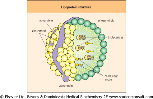
| A lipoprotein particle contains a hydrophobic core of cholesterol esters and triacylglycerols (Fig. 17.2). Amphipathic phospholipids and free cholesterol, together with apoproteins form an outer layer. Some proteins, such as apoprotein B (apoB), are embedded in the particles. Others, such as apoC, are only loosely bound - and can be easily exchanged with other particles.
|
| ULTRACENTRIFUGATION IS A POWERFUL SEPARATION METHOD |
In the clinical laboratories simple centrifuges are used to separate serum (or plasma) from red blood cells. These machines employ a moderate centrifugal force, 2000-3000 g to sediment the blood cells. However, when much larger centrifugal forces (40 000-100 000 g) are applied to plasma, centrifugation, now termed ultracentrifugation, becomes a powerful separation method for particles and molecules. Ultracentrifugation is extensively used in lipid research. When the centrifugal force is applied to a solution, particles that are heavier than the surrounding solvent sediment, and those lighter than the solvent float to the surface at a rate proportional to the applied centrifugal force and to the particle size. The formula below summarizes factors that affect the particle movement:
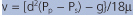 where v = sedimentation rate, d = diameter, Pp = particle density, Ps = solvent density, μ = viscosity of the solvent, and g = gravitational force.
 |
| In a technique known as flotation ultracentrifugation, plasma containing lipoproteins are overlayered with a solution of defined density, e.g. 1.063 kg/L, the density of VLDL. After several hours of centrifugation (depending on the type of centrifuge rotor, the rotor speeds would be in the range of 40 000 rev/min), the VLDL float to the surface, where they can be harvested. Other density solutions can be used to separate other lipoproteins. Variations of the ultracentrifugation technique, such as density gradient centrifugation, can be applied to separate a plasma sample into several 'bands' containing different lipoprotein fractions. Ultracentrifugation is also extensively used in protein and nucleic acid biochemistry. |
| LABORATORY TESTING FOR LIPID DISORDERS |
| There are several 'levels' of lipid testing in clinical situations |
| There are three stages of testing in the diagnosis of lipoprotein disorders (Fig. 17.5A). The first stage is the screening for just total cholesterol. This can be done on either a fasting or nonfasting sample because cholesterol changes little during the fast-fed cycle. The second stage consists of measurements of total cholesterol, triacylglycerol (triglycerides), and HDL-cholesterol in serum. Such measurements need to be performed after a 12-h fast, because triacylglycerol concentration increases postprandially. The LDL concentration is usually calculated from the values of total cholesterol and HDL-cholesterol (Fig. 17.5B). The final step performed in specialist laboratories is lipoprotein analysis by ultracentrifugation. |
| page 226 |  | | page 227 |
|
Table 17-1.
The characteristics of the main lipoprotein classes. |
| Body_ID: None |
| The lipoprotein classes |
| Body_ID: T017001.50 |
| Particle | Density (kg/L) | Main component | Apoproteins* | Diameter (μm) |
| Body_ID: T017001.100 |
| chylomicrons | <0.95 | TG | B48 (A, C, E) | 75-1200 |
| Body_ID: T017001.150 |
| VLDL | 0.95-1.006 | TG | B100 (A, C, E) | 30-80 |
| Body_ID: T017001.200 |
| IDL | 1.006-1.019 | TG & cholesterol | B100, E | 25-35 |
| Body_ID: T017001.250 |
| LDL | 1.019-1.063 | Cholesterol | B100 | 18-25 |
| Body_ID: T017001.300 |
| HDL | 1.063-1.210 | Protein | AI, AII (C, E) | 5-12 |
| Body_ID: T017001.350 |
|
| Body_ID: T017001.400 |
*Main apoproteins present in a given lipoprotein particle are indicated first, with those that are exchanged with other particles in brackets.
TG, triacylglycerol (triglyceride); VLDL, very-low-density lipoproteins; IDL, intermediate-density lipoproteins; HDL, high-density lipoproteins. When separated by electrophoresis VLDL are called pre-beta lipoproteins, LDL, beta lipoproteins and HDL alpha lipoproteins.
|
| Figure 17.2 The lipoprotein particle. The external monolayer of a lipoprotein particle contains free cholesterol, phospholipids, and apoproteins. Cholesterol esters and triacylglycerols locate in the particle core. |
| Lipoprotein particles present in plasma form a continuum of size and density (Table 17.1). Their classification is based on their hydrated density. Thus there are chylomicrons, very-low-density lipoproteins (VLDL), remnant particles (which include intermediate-density lipoproteins [IDL]), LDL, and high-density lipoproteins (HDL). VLDL and remnant particles are triacylglycerol-rich, whereas LDL is cholesterol-rich.
The density of these particles increases and the size decreases with decreasing triacylglycerol content, from chylomicrons (the lightest) through VLDL, remnant particles, IDL, LDL, to HDL (the heaviest). HDL contains several proteins, and some cholesterol and phospholipids, but relatively little triacylglycerol.
|
| One can also characterize lipoproteins by their electrophoretic mobility
|
| MUTATION IN APOPROTEIN E GENE CAUSES DYSLIPIDEMIA INVOLVING REMNANT PARTICLES |
| Apoprotein E plays a key role in the metabolism of lipoprotein remnants. Its synthesis is controled by three major alleles, e2, e3, and e4. It is a relatively small protein, with a molecular mass of 34 kDa. There are three major isoforms: E2, E3, and E4. ApoE is recognized both by the LDL (apo B/E) receptor and by the LRP. The E2 ioform has much lower affinity (1%) to the receptor than other isoforms. This, in homozygotes, slows down the uptake of remnants and results in familial dyslipidemia (also known as type III 3 hyperlipidemia). Also, apoE seems to be important for lipid metabolism in the nervous system where it is synthesized by brain astrocytes. The presence of an e4 allele is associated with Alzheimer's disease, a disorder responsible for at least 50% of all cases of dementia. |
| On electrophoresis α-lipoproteins (HDL) migrate furthest towards the anode (+electrode), followed by pre-β-lipoproteins (VLDL) and β-lipoproteins (LDL). Chylomicrons remain at the cathodic end, at the origin of the electrophoretic strip. The electrophoretic pattern is the basis for
the phenotypic classification of dyslipidemias into five types adopted by the WHO (Table 17.2). However, when it became evident that a disorder of lipid metabolism may present with different phenotypic patterns (see Table 17.3) dyslipidemias were re-classified; they are now defined either as separate genetically-determined entities (e.g. familial hypercholesterolemia) or simply classed according to plasma levels of cholesterol, triacylglycerol, and HDL-cholesterol (see below).
|
|