Potential Hydrogen Bonding Patterns by Proteins to DNA Base Pairs

Overlay with: GC TA AT Numbering.

Overlay with: AT CG GC Numbering.
Potential Hydrogen Bonding Patterns by Proteins to DNA Base Pairs | |
|---|---|
| CG Base Pair | TA Base Pair |
 Overlay with: GC TA AT Numbering. |  Overlay with: AT CG GC Numbering. |
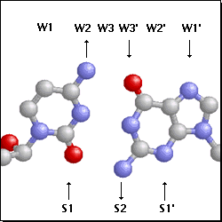
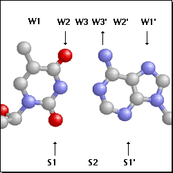
The CG and TA base pairs are shown as Ball & Stick with CPK coloring. Potential
H-bond interactions with protein residues in the major groove are shown at the top;
interactions in the minor groove are at the bottom. Arrows indicate the H-bond donor
--> acceptor direction.
The effects of a base pair substitution on these potential H-bond interactions can be seen
by selecting the base pair overlays under each image. The dark gray Stick diagrams of each
substitution are colored (CPK) at the positions in both grooves where H-bonding could
occur. (The 5-methyl group of T is colored black; Watson-Crick H-bonding is not shown.)
If the base pair substitution results in a loss of a H-bond, the orignal interaction is
colored red. If the substitution causes only a shift in position, that interaction is
colored orange. For example, the substitution of CG with TA would lose potential H-bonds
at positions W2, W3', and S2, but leave those at W1', S1,
and S1' intact.
Also note that the positions, W1, S1, etc. are highly diagramatic;
they are intended to serve only as constant markers for the different base pairs, not as
fixed positions for real protein residues.
View two examples of actual H-bonding in the Eco RI-DNA Complex.
Adapted from the figures and descriptions in Seeman, N. C. et al. (1976) "Sequence-specific recognition of double helical nucleic acids by proteins"Proc. Natl. Acad Sci.73, 804.