 Back to the Chaperonin Page
 Email comments or suggestions to Jeff Seale
Last Updated December 26, 1997
Structural Features of the Chaperonins
The structure of a GroEL monomer taken from the original
structure determination (335 Kb)
The refined x-ray structure of a GroEL heptamer (2.25 Mb)
The x-ray structure of a GroEL 14mer complexed with ATP-gamma-S
(5.8 Mb)
The x-ray structure of a GroEL14-ADP7-GroES7 asymmetric complex
(4.99 Mb)
The x-ray structure of a GroEL apical domain fragment (110
kb)
The x-ray structure of a GroEL apical domain fragment double
mutant A262L, I267M (162 kb)
WARNING: These PDB files are large. Viewing them through your Web browser may be VERY slow. In order to best view these files, you might want to save them to your disk and view them from your hard drive.
To view PDB files, download free Rasmol software here
An Overview of the GroEL and GroES Structures can be found here
This page was created by Will McClure as part of the Physical Biochemistry Course at Carnegie Melon. There are nice pictures of GroEL highlighting residues involved in GroES binding, polypeptide binding and ATP binding.
Gif files of the Cpn10 from Mycobacterium leprae courtesy of Christophe Verlinde.
For a reference, see: Mande, et al. Science 271: 203-207 (1996)
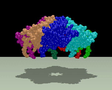
A space filling model of the Cpn10 heptamer .
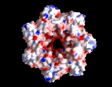
An electrostatic potential surface viewed from inside the
heptamer highlighting the negative charge around the roof.
Cryo-EM Images from Helen Saibil's Chaperone Page at
Birkbeck
Movies of the ATPase cycle from Birkbeck.
Images from Jose Valpuesta's
Group in Spain
Images of symmetric
GroEL-GroES complexes with two rhodanese bound from Jose Valpuesta's Group in Spain
Back to the Chaperonin Page
Email comments or suggestions to Jeff Seale
Last Updated December 26, 1997