| TYPES OF TRANSPORT PROCESSES
|
| Simple diffusion through the phospholipid bilayer
|
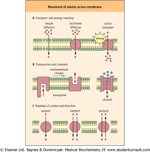
Small, nonpolar molecules (such as O2, CO2, N2) and uncharged polar molecules (such as urea , ethanol, and small organic acids) move through membranes by simple diffusion without the aid of membrane proteins (Table 7.3 and Fig 7.3A). The direction of net movement of these species is always 'downhill', along the concentration gradient, from high to low concentration to establish equilibrium. , ethanol, and small organic acids) move through membranes by simple diffusion without the aid of membrane proteins (Table 7.3 and Fig 7.3A). The direction of net movement of these species is always 'downhill', along the concentration gradient, from high to low concentration to establish equilibrium.
|
| The hydrophobicity of the molecules is an important requirement for simple diffusion across the membrane, as the interior of the phospholipid bilayer is hydrophobic. The rate of transport of a small molecule is, in fact, closely related to its partition coefficient between oil and water.
|
| page 80 |  | | page 81 |
| EXTRACTION AND ANALYSIS OF MEMBRANE PHOSPHOLIPIDS |
| Cells (2 × 10⁷) or membranes (2 mg protein) are suspended in phosphate-buffered saline (1.6 ml) in a screw-capped glass test tube. Chloroform/methanol (1:2, v/v) (6 ml) is added into the tube and lipids are extracted by vigorous mixing. Equal volume of chloroform and the saline (2 ml each) are further added and again mixed. After centrifugation at 1,500 × g for 5 min, the lower phase (chloroform containing extracted lipids) is recovered, and the solvent is evaporated under the stream of N2 gas. The lipid extract is applied to silica gel plate for two-dimensional thin-layer chromatography with chloroform/methanol/acetic acid (65:25:10, v/v) as the solvent in the first dimension, and chloroform/methanol/formic acid (65:25:10, v/v) in the second. The separated phospholipids are detectable on the plate as yellow spots by iodine vapor. Using appropriate standards, the ratio of lecithin (phosphatidylcholine) to sphingomyelin, measured in amniotic fluid, can be used to assess fetal lung maturity and risk for acute respiratory distress syndrome (ARDS, Chapter 26). |
| Figure 7.3 Various models of movement of solute across membranes. |
|
Table 7-3.
Classification of transport systems of biomembranes. |
| Body_ID: None |
| Transport systems of biomembranes |
| Body_ID: T007003.50 |
| Type | | Transport protein (example) | Energy coupling | Specificity | Saturability | Rate (molecules/transport protein/s) |
| Body_ID: T007003.100 |
| Passive transport or diffusion | simple diffusion facilitated diffusion
transporter
channel | -
+
(GLUT-1∼5)
(H2O, Na+, K+, Ca2+, Cl-) | -
- | -
+ | -
+ |
-102
107-108 |
| Body_ID: T007003.150 |
| Active transport | primary secondary
symporter
antiporter
uniporter | +
(see Table 7.5)
+ (SGLT-1, 2, neutral amino acids)
(Cl-/HCO3-, Na+/Ca2+, Na+/H+)
(Glutamate) | +
+ | +
+ | +
+ | 102-104
100-102* |
| Body_ID: T007003.200 |
|
| Body_ID: T007003.250 |
*The Cl-/HCO3- antiporter seems to be an exception to secondary active transport systems, as its transport rate is high, at 105 mol/s.
Transport systems are classified according to the role of transport proteins and energy coupling. Typical substrates for various types of channels are shown in the parentheses.
|
| page 81 | | | page 82 |
| Although the fluid mosaic model is basically correct, it is recognized that there are membrane patches with unique protein and lipid compositions separated from other fluid membrane areas. Caveolae, 50-100 nm plasma membrane invaginations, and lipid rafts are plasma membrane patches (microdomains) important for signal transduction through immune and growth factor receptors. These patches are enriched in cholesterol and sphingolipids, and the interaction of the long saturated fatty acid tails of sphingolipids with cholesterol results in the stabilization of their less fluid environment. The patches are detergent-insoluble and show high buoyant density on sucrose density gradient centrifugation. While the patches are associated with specific molecules such as GPI-anchored proteins, caveolins are the principal components of caveolae. Caveolae are believed to be involved in cholesterol transport, the transport of solutes across endothelial cells, and tumor suppression. Pathogens such as viruses, parasites, bacteria and even bacterial toxins enter into the host cells through caveolae. It is interesting that the sites of γ-secretase activity and Aβ production, which are closely related to the pathology of Alzheimer's disease, are associated with membrane regions of high cholesterol content, such as lipid rafts. |
| Examples of patches enriched in a particular protein are the purple membrane of Halobacterium halobium containing bacteriorodopsin, and gap junctions containing connexin. Bacteriorodopsin is a light driven-proton pump which generates H+-concentration gradient across the bacterial membrane. The gradient is used for ATP synthesis and nutrient uptake for bacterial growth. |
| Although water molecules can be transported by simple diffusion, channel proteins are believed to control the movement of water across most membranes, especially in the kidney for concentration of the urine. Mutation in a water
channel protein gene (aquaporin-2) causes diuresis in some patients with nephrogenic diabetes insipidus.
|
|