| Xeroderma Pigmentosum (XP) is a group of rare, life-threatening, autosomal recessive disorders (incidence = 1/250 000) that are marked by extreme sensitivity to sunlight. Upon exposure to sunlight or ultraviolet radiation, the skin of XP patients erupts into pigmented spots, resembling freckles. Multiple carcinomas and melanomas appear early in life, exacerbated by sun exposure, and the majority of patients succumb to cancer before reaching adulthood. |
| XP is the result of a defect in repair of UV-induced thymine dimers in DNA. There are at least eight genes that are involved in repair of UV-induced thymine dimers. Patients with XP must avoid direct sunlight, fluorescent light, halogen light, or any other source of ultraviolet light. An experimental form of protein therapy, currently undergoing clinical evaluation, involves application of a skin lotion containing the missing protein or enzyme. Ideally, this protein will enter the skin cells and stimulate the repair of UV damaged DNA. However, protection occurs only where the lotion can be applied. For example, this treatment does not address the neurological problems that affect about 20% of XP patients. |
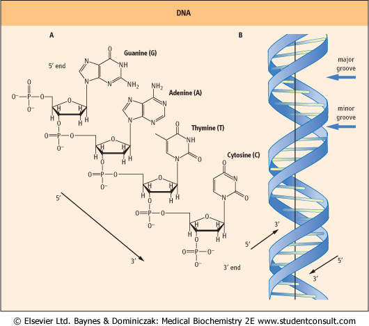
| DNA is an anti-parallel dimer of nucleic acid strands. It is composed of nucleotides containing the sugar deoxyribose. Deoxyribose is missing the hydroxyl group at the 2'-position. The chains of DNA are polymerized through a phosphodiester linkage from the 3'-hydroxyl of one subunit to the 5'-hydroxyl of the next subunit (Fig. 30.1A). Thus, DNA is a
linear deoxyribose phosphate chain with purine and pyrimidine bases attached to carbon-1 of the deoxyribose subunit.
|
| Using X-ray diffraction photographs of DNA taken by Rosalind Franklin, a structure for DNA was proposed by James Watson and Francis Crick in 1953. This model proposed that DNA was composed of two intertwined complementary strands with hydrogen bonds holding the strands together (Fig. 30.1B). The basic simplicity of this structure led to its rapid acceptance. While some of the details of the model have been modified, its essential elements have remained unchanged since originally proposed.
|
| Watson and Crick model of DNA
|
| page 425 |  | | page 426 |
| Figure 30.1 Structure of DNA. (A) A tetranucleotide sequence of DNA showing each of the nucleotides normally found in DNA. The deoxyribose sugars are missing the 2'-hydroxyl that is present in the ribose sugars found in RNA. By convention, DNA is read from the 5' to 3' end, so the sequence of this tetranucleotide is 5'-GATC-3'. (B) A graphic representation of the structure of B-DNA, the major form of DNA in the cell. The base pairs in the middle are aligned nearly perpendicular to the helical axis. The major groove and the minor groove are shown. Note that the strands are antiparallel. |
| As originally presented by Watson and Crick, DNA is composed of two strands, wound around each other in a right-handed, helical structure with the base pairs in the middle and the deoxyribosylphosphate chains on the outside. The orientation of the DNA strands is anti-parallel (i.e. the strands run in opposite directions). The nucleotide bases on each
strand interact with the nucleotide bases on the other strand to form base pairs (Fig. 30.2). The base pairs are planar and are oriented nearly perpendicular to the axis of the helix. Each base pair is formed by hydrogen bonding between a purine and a pyrimidine. Guanine forms three hydrogen bonds with cytosine, and adenine forms two with thymine. Because of the specificity of this interaction between purines and pyrimidines on the opposite strands, the opposing strands of DNA are said to have complementary structures. The composite strength of the numerous hydrogen bonds formed between the bases of the opposite strands is responsible for the extreme stability of the DNA double helix. While the hydrogen bonds between strands are affected by temperature and ionic strength, stable complementary structures can be formed at room temperature with as few as six to eight nucleotides.
|
| The three-dimensional structure of the DNA double helix is such that the deoxyribosyl phosphate backbones of the two strands are slightly offset from the center of the helix. Because of this, the grooves between the two strands are of different sizes. These grooves are termed the major groove and the minor groove (see Figs 30.1B and 30.2). The major groove is more open and exposes the nucleotide base pairs. The minor groove is more constricted, being partially blocked by the deoxyribosyl moieties linking the base pairs. Binding of proteins to DNA frequently occurs in the major groove and is specific for the nucleotide sequence of DNA.
|
|