| Figure 34.13 RFLP analysis in sickle-cell anemia. An A > T substitution at codon 6 of the β-globin gene abolishes a recognition site for the enzymes, MstII, which can be used to determine the presence or absence of the mutation by studying the RFLP pattern. |
|
Table 34-5.
Applications of PCR. |
| Body_ID: None |
| Applications of PCR |
| Body_ID: T034005.50 |
| Application |
| Body_ID: T034005.100 |
| Genetic marker typing | restriction site polymorphisms |
| Body_ID: T034005.150 |
| | microsatellite repeats |
| Body_ID: T034005.200 |
| Detection of point mutations | restriction site polymorphisms |
| Body_ID: T034005.250 |
| | amplification refractory mutation systems |
| Body_ID: T034005.300 |
| Amplification of | double-stranded DNA |
| Body_ID: T034005.350 |
| DNA templates for | single-stranded templates |
| Body_ID: T034005.400 |
| DNA sequencing | |
| Body_ID: T034005.450 |
| Genomic DNA cloning | PCR of gene families or genes in different species |
| Body_ID: T034005.500 |
| Others | genome walking, introduction of mutations in vitro to test their effect in biological systems |
| Body_ID: T034005.550 |
| PCR is a versatile method applied to a multitude of methods in DNA research (Table 34.5). One of the most widely used clinical applications of PCR is in the identification of
genetic mutations within PCR amplified DNA from 'at risk' individuals.
|
| Assay of restriction site polymorphisms
|
| Restriction site polymorphisms (RSPs) are small-scale equivalents of RFLPs
|
| Naturally occurring variations in DNA sequence may abolish or create restriction sites that can be detected conveniently by digesting PCR amplified sequences with the appropriate restriction enzyme.
|
| page 487 |  | | page 488 |
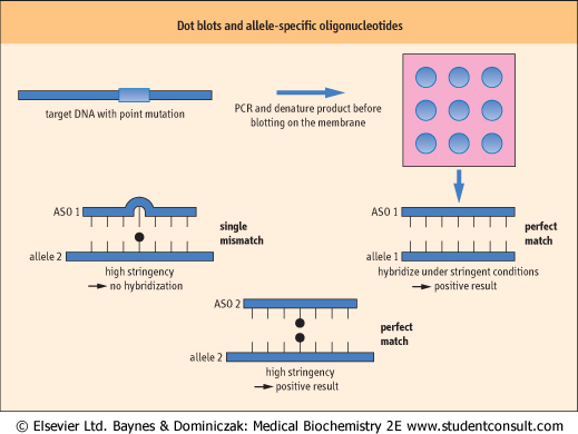
| Figure 34.14 Dot blots and allele-specific oligonucleotides. Using a technique similar to Southern blotting, single-stranded PCR products are transferred onto a nylon membrane and fixed in situ. Allele-specific oligonucleotides (ASOs) are hybridized to the target DNA under conditions of high stringency. In this case, the ASOs are used to identify a single base substitution, with one detecting the wild type and another the mutant allele. |
| Detection of microsatellite repeats
|
| This technique detects sets of tandem nucleotide repeats present in mammalian genomes
|
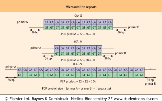
| Tandem microsatellite repeats are sequences that comprise a few nucleotides - between one and four but typically two or three, e.g. CA or CAG, that are repeated in tandem at various locations throughout mammalian genomes, so-called microsatellite repeats. These microsatellites are small blocks of DNA that occur in non-coding regions, either intergenic or within introns of genes. The term 'satellite DNA' has been historically applied to regions of repetitive sequence in which the local base composition may differ from the overall composition of human DNA. One important feature of these microsatellite repeats is that they are highly polymorphic, i.e. the number of tandem repeats in a particular microsatellite may vary from one individual to another. This means that the two copies of a single human microsatellite, one from each chromosome, may be of different lengths and can thus be distinguished in that individual. The number of different alleles of the microsatellite will vary depending on which microsatellite is studied, but may vary from 2-15 or more copies of the repeat per allele (Fig. 34.15). As a result of this high degree of polymorphism, microsatellite repeats are of great value in the study of genetic linkage. This is because a particular microsatellite allele may fortuitously be located in the vicinity of a pathological mutation. Comparison of the DNA of affected families may indicate that a particular microsatellite serves as a marker with which to trace the presence or inheritance of the mutation under study.
|
| Mutation detection by allele-specific PCR
|
| Only mutant DNA will be amplified using this technique
|
| Standard PCR employs two primers to amplify both strands of the DNA and relies on the fact that each primer will reliably amplify the target strand. However, PCR can be performed using allele-specific primers, which will only amplify one allele of a target gene. This means that if a primer contains a sequence that recognizes a mutation, it will only hybridize with the mutant DNA. Thus, only DNA containing the mutation will amplify with this primer. The allele-specific primer has its extreme 3'-nucleotides as the ones that detect the mutation, as the DNA polymerase requires complete hybridization of primer and template at the 3' end of the primer. This method can be applied to the detection of specific mutations in a single gene, e.g. the gene for cystic fibrosis. This type of PCR is often called an amplification refractory mutation system (ARMS) as PCR amplification will be refractory, i.e. will not occur, in the absence of the mutant allele.
|
| page 488 | | | page 489 |
| Figure 34.15 Microsatellite repeats. A typical microsatellite contains tandem repeats of a dinucleotide, e.g. CA. The number of repeats can vary from 2 to 20 or more. Amplification of the DNA including the microsatellite repeat is performed by PCR using a radiolabeled primer and the products are then size-fractionated on a polyacrylamide gel to aid resolution. PCR product size = 72 (primer A + primer B) +26 (13 × 2 for each dinucleotide) = 98. In this instance, three alleles can be identified: (CA)13, (CA)9, and (CA)24. |
| MICROSATELLITE REPEATS ARE IDEAL MARKERS FOR STUDY OF INHERITANCE |
| A 7-year-old boy was the subject of disputed paternity. His mother had identified a man as the father and maintained that he should be responsible for the child's financial well-being. All parties consented to DNA testing by means of DNA fingerprinting. Study of the DNA tests revealed that this man was the father and was indeed responsible for the child. |
| Comment. The most commonly studied microsatellites contain dinucleotide repeats of (CA) or (TG) and may vary in repeat number from one or 2, up to 20 or more. Thus, a standard dinucleotide repeat would be denoted (CA)n, where n is the number of repeats. An ideal microsatellite marker would have a large number of alleles, i.e. n > 100, and each allele would be equally frequent. However, most microsatellites have less than 20, and there is often a preponderance of one or two particular alleles in any population. When microsatellites are typed by PCR amplification and gel electrophoresis, both chromosomes are amplified and thus two possible alleles are generated from each locus for a single individual. |
| In forensic medicine, simple questions are often asked, e.g. 'Is this man the child's father?' or 'Does this blood stain correspond to blood of the suspect?' By selecting several microsatellite markers, typically six to nine, that are highly polymorphic and have an even spread of allele frequency, i.e. each allele is equally frequent, a series of microsatellite markers can be generated. When an individual's markers are tested, the result is a set of alleles that are virtually unique to an individual, a so-called 'DNA fingerprint'. Thus, by comparing the pattern of microsatellites generated in the subject in question and comparing this with the pathologic sample or the DNA of offspring, the identity of a parent or suspect can be confirmed or refuted with a high degree of certainty. |
| Microsatellites have also been used to examine the linkage of a disease trait, e.g. type 1 diabetes, to certain regions of the human genome. By studying the inheritance of microsatellite markers in families or the sharing of alleles in affected siblings, a statistical measure of the degree of co-inheritance of a marker can be determined. This approach has been used successfully to identify genetic loci strongly linked to the inheritance of type 1 and type 2 diabetes, and locate the genes causing Huntington's disease, myotonic dystrophy and other single gene disorders (Table 34.6). |
| page 489 | | | page 490 |
|
Table 34-6.
Unstable trinucleotide repeats. |
| Body_ID: None |
| Unstable trinucleotide repeats |
| Body_ID: T034006.50 |
| Disease | Repeat | Normal Length | Mutation length |
| Body_ID: T034006.100 |
| Huntington's disease | (CAG)n | A:9-35 | 37-100 |
| Body_ID: T034006.150 |
| Kennedy disease | (CAG)n | 17-24 | 40-55 |
| Body_ID: T034006.200 |
| Spinocerebellar ataxia I (SCA I) | (CAG)n | 19-36 | 43-81 |
| Body_ID: T034006.250 |
| Dentatopallidoluysianatrophy (DRPLA) | (CAG)n | 7-23 | 49-75+ |
| Body_ID: T034006.300 |
| Machado-Josephdisease (SCA III) | (CAG)n | 12-36 | 69-79+ |
| Body_ID: T034006.350 |
| Fragile X site A (FRAXA) | (CGG)n | 6-54 | 200-1000+ |
| Body_ID: T034006.400 |
| Fragile X site E (FRAXE) | (CCG)n | 6-25 | >200 |
| Body_ID: T034006.450 |
| Fragile X site F (FRAXF) | (GCC)n | 6-29 | >500 |
| Body_ID: T034006.500 |
| Fragile 16 site A (FRA16A) | (CCG)n | 16-49 | 1000-2000 |
| Body_ID: T034006.550 |
| Myotonic dystrophy | (CTG)n | 5-35 | 50-4000 |
| Body_ID: T034006.600 |
| Friedreich ataxia | (GAA)n | 7-22 | >200 |
| Body_ID: T034006.650 |
|
| Body_ID: T034006.700 |
In all of the cases listed, trinucleotide repeats are present in individuals without clinical disease. However, progressive increase in the number of repeats leads to the onset of clinical signs, although a premutant phase has been described in some disorders, such as FRAXA, where expansions larger than normal are found but with minimal or no clinical features. These premutant states give rise to carriers - females who can subsequently pass on an expanded triplet repeat to their offspring, which may result in the disease.
|
| Detection of deletions in genes causing disease
|
| This is performed using primers that generate PCR products of different sizes
|
| In patients with Duchenne muscular dystrophy, many of the disease-causing mutations are due to deletions of one of relatively few exons in this, one of the largest of human genes. Specific amplification of these exons, using primers that generate PCR products of different sizes for each exon, can be used to screen quickly for the presence of a disease-causing deletion in an affected individual or determine if the fetus of a carrier has the deletion. Such PCR is carried out in a single reaction using a single template and 5-10 different primer pairs, each generating a separate amplification product. This form of PCR is called multiplex PCR.
|
|