| DAG activates protein kinase C
|
| page 550 |  | | page 551 |
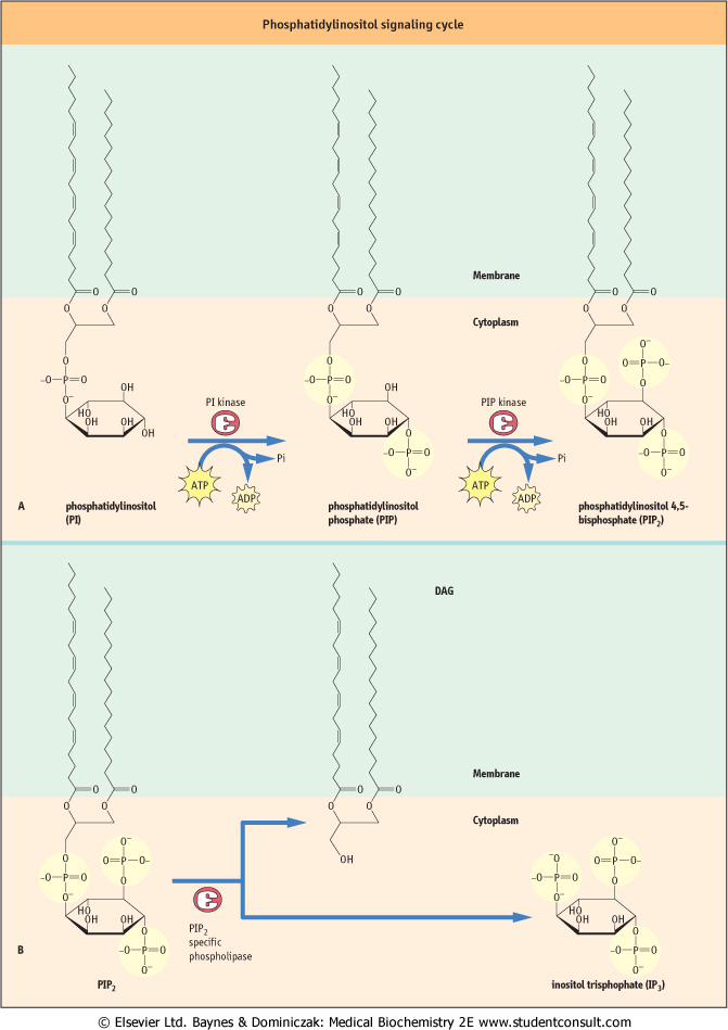
| Figure 38.8 The phosphatidylinositol signaling cycle. |
| page 551 | | | page 552 |
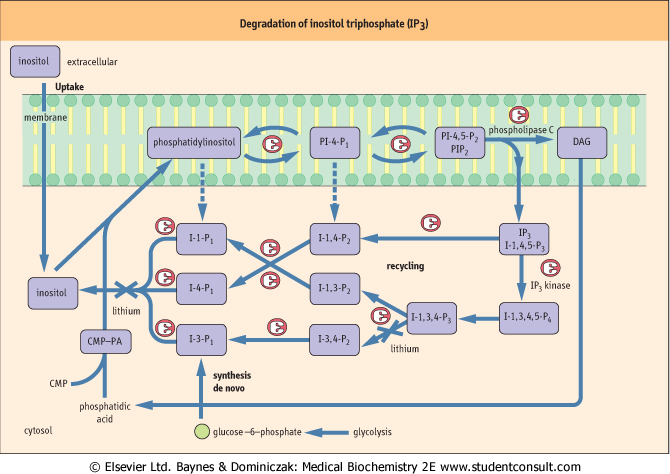
| Figure 38.9 Degradation of inositol triphosphate (IP3). The major pathways involve (i) the sequential action of phosphatases converting I-1,4,5-P3 to inositol, and (ii) an IP3 kinase, which generates I-1,3,4,5-P4, which is in turn sequentially degraded to inositol by inositol phosphate-specific phosphatases. DAG, diacylglycerol; PA, phosphatidic acid; CMP-PA, cytosine monophosphate-phosphatidic acid. |
|
Table 38-3.
The protein kinase C (PKC) superfamily. |
| Body_ID: None |
| Protein kinase C superfamily |
| Body_ID: T038003.50 |
| | Classical PKCs | | Novel PKCs | | | Atypical PKCs | | |
| Body_ID: T038003.100 |
| PKC designation | α | β1 | β2 | γ | δ | ε/ε' | η | [thetas] | μ | ι/λ | ζ |
| Body_ID: T038003.150 |
| molecular mass (kDA)activators | 82 | 80 | 80 | 80 | 78 | 90 | 80 | 79 | 115 | 74 | 72 |
| Body_ID: T038003.200 |
| [Ca2+] | yes | yes | yes | yes | no | no | no | no | | no | no |
| Body_ID: T038003.250 |
| DAG | yes | yes | yes | yes | yes | yes | yes | yes | | no | no |
| Body_ID: T038003.300 |
| tissue distribution | all | some | many | neural | all | neural | many | muscle, skin | | ovary, testis | all |
| Body_ID: T038003.350 |
|
| Body_ID: T038003.400 |
This is a multigene family, except for β1, β2 and ε/ε', which are alternatively spliced forms of β and ε. DAG, diacylglycerol.
|
| page 552 | | | page 553 |
| DAG fulfils its second messenger role by activating the key signaling enzyme, protein kinase C (PKC), which phosphorylates a wide range of target signal-transduction proteins on serine or threonine. PKC was originally identified as a calcium- and lipid (phosphatidylserine)-dependent kinase important in the regulation of cell proliferation. However, in recent years it has become clear that PKC is, in reality, a generic name for a superfamily of related kinases that have different tissue distribution and activation requirements (Table 38.3). Nevertheless, all these enzymes share some conserved features;
most notably they comprise two major domains: an N-terminal regulatory domain and a C-terminal catalytic kinase domain. The regulatory domain contains a pseudosubstrate sequence that resembles the consensus phosphorylation site in PKC substrates. In the absence of activating cofactors (Ca2+, phospholipid, DAG), this pseudosubstrate sequence interacts with the substrate-binding pocket in the catalytic domain and represses PKC activity; binding of cofactors reduces the affinity of this interaction, induces a conformational change in the PKC, and allows stimulation of PKC activity. Consistent with the fact that the activator/cofactor, DAG, is anchored in the membranes, PKC activation is
generally associated with translocation from the cytosol to the plasma or nuclear membranes. Although PKC is generally considered to be a serine kinase, it can phosphorylate threonine, but never tyrosine, residues. Interestingly, PKC can phosphorylate the same protein targets as PKA but, whereas PKC generally phosphorylates the protein at serine residues, PKA usually phosphorylates threonine residues.
|
|