| CHARACTERISTICS OF MAMMALIAN GLOBIN PROTEINS
|
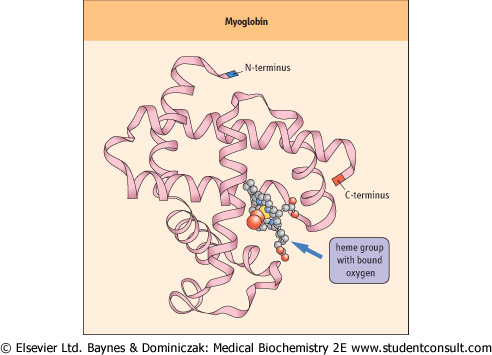
Globins constitute an ancient family of soluble metalloproteins whose structure and function have been preserved for several million years among leguminous plants, certain invertebrates, and vertebrates. These proteins most likely evolved to convert heme from an electron carrier to an oxygen carrier. Mb and Hb are examples of globins found in all mammals. Mb consists of a single globin polypeptide and a heme prosthetic group (Fig. 4.2). Hb is a tetrameric assembly of closely related globin subunits. The globin polypeptide is a single chain of approximately 150 amino acids . Each globin molecule contains one noncovalently bound heme prosthetic group. The most significant aspect of the secondary structure of globins is the high proportion of α-helix: over 75% of the amino acids are associated with eight helical segments containing as few as six and as many as 28 residues. These α-helices are organized into a tightly packed, nearly spherical, globular tertiary structure (Fig. 4.2). . Each globin molecule contains one noncovalently bound heme prosthetic group. The most significant aspect of the secondary structure of globins is the high proportion of α-helix: over 75% of the amino acids are associated with eight helical segments containing as few as six and as many as 28 residues. These α-helices are organized into a tightly packed, nearly spherical, globular tertiary structure (Fig. 4.2).
|
| Figure 4.2 Model of myoglobin. Myoglobin is a compact globular protein. In this depiction of mammalian Mb only the globin polypeptide backbone is shown, with emphasis on the high proportion of secondary structure (exclusively α-helix). The N-terminus is blue; the C-terminus is red. The heme group, with bound O2, is illustrated as a 'ball-and-stick' structure. |
| page 36 |  | | page 37 |
| HYPERBARIC O2 THERAPY IN TREATMENT OF ACUTE CO POISONING |
| A 22-year-old pregnant female, carrying a fetus of 31 weeks gestational age, was transported to the maternity clinic of a hospital for suspected CO poisoning. The patient was experiencing headache, nausea, and visual abnormalities. She stated that her workplace had been undergoing repairs to the heating and ventilation systems during the past 2 weeks, and on the day of her hospital visit the fire department had evacuated the building after detecting a high level of CO (200 ppm). Vital signs were blood pressure of 116/68 mm Hg, pulse rate of 100, and respiratory rate of 24. Noteworthy in the patient's evaluation was a carboxyhemoglobin component of 15% of total Hb at time of admission (normal <3%, but may exceed 10% in heavy smokers). External fetal monitoring indicated a fetal heart rate of 135, with occasional, moderate irregularities. Uterine contractions were occurring every 3-5 min. The patient was treated in the hospital's hyperbaric O2 chamber: 30 min at 250 kPa (2.5 atmospheres), then 60 min at 200 kPa. She also received magnesium sulfate intravenously to resolve the premature contractions. The patient was discharged 2 days later. She delivered a healthy female infant at 38 weeks of gestational age who, on examination at birth and at 6 weeks of age, exhibited no apparent sequelae to her in utero exposure to CO. |
| Comment. Like O2, CO binds to heme prosthetic groups. Because the affinity of globin-bound heme for CO is more than 104 times that of O2, prolonged exposure of hemoglobin (Hb) to CO would be virtually irreversible (t1/2 = 4-5 h) and lead to highly toxic levels of carboxyhemoglobin. Hyperbaric O2 is the treatment of choice for severe or complicated CO poisoning. The administration of 100% O2 at 200-300 kPa creates arterial and tissue pO2 values of 2000 mmHg and 400 mmHg respectively (∼20 times normal). The immediate result is a reduction in the t1/2 of carboxyhemoglobin to less than 20 min. Hyperbaric O2 is also used in the treatment of decompression sickness, arterial gas embolism, radiation-induced or ischemic tissue injury, and severe hemorrhage. |
|
| Polar amino acids are located almost exclusively on the exterior surface of globin polypeptides and contribute to the remarkably high solubility of these proteins (for example, 5.2 mmol/L [335 g/L] Hb in the erythrocyte, or >30% protein). Amino acids that are both polar and hydrophobic,
such as threonine, tyrosine, and tryptophan, are oriented with their polar functions toward the protein's exterior. Hydrophobic residues are buried within the interior, where they stabilize the folding of the polypeptide and form a pocket that accommodates the heme prosthetic group. Notable exceptions to this general distribution of amino acid residues in globins are the two histidines that play indispensable roles in the heme pocket. The side chains of these histidines are oriented perpendicular to and on either side of the planar heme prosthetic group. One histidine has an imidazole nitrogen that is close enough to bond directly to the Fe2+ atom: this is the proximal histidine. On the opposite side of the heme plane is the other histidine: this is the distal histidine. The distal histidine is too far from the heme iron for direct bonding; rather, it confers important geometrical constraints on the sixth coordination site and makes a hydrogen bond with the bound O2. The alignment of heme and the distal histidine permits O2 to bind favorably to the Fe2+ atom.
|
|