| The interconversion stage of the pentose phosphate pathway
|
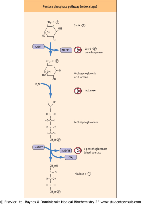
| Figure 11.9 The redox stage of the pentose phosphate pathway. A sequence of three enzymes forms 2 moles of NADPH per mole of Glc-6-P, which is converted into ribulose-5-phosphate, with evolution of CO2. |
| page 151 |  | | page 152 |
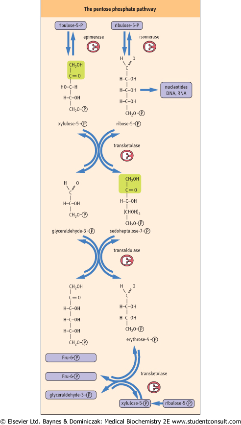
| Figure 11.10 The interconversion stage of the pentose phosphate pathway. The carbon skeletons of three molecules of ribulose-5-phosphate are shuffled to form two molecules of Fru-6-P and one molecule of glyceraldehyde 3-phosphate. |
|
Table 11-2.
Summary of equilibrium reactions in the Pentose Phosphate Pathway. |
| Body_ID: None |
| The Pentose Phosphate Pathway |
| Body_ID: T011002.50 |
| Substrate(s) | | Product(s) | Enzyme |
| Body_ID: T011002.100 |
| Ribulose-5-P | | Ribose-5-P | isomerase |
| Body_ID: T011002.150 |
| 2 Ribulose-5-P | | 2 Xylulose-5-P | epimerase |
| Body_ID: T011002.200 |
| Xylulose-5-P | | Glyceraldehyde-3-P | transketolase |
| Body_ID: T011002.250 |
| + Ribose-5-P | | + Sedoheptulose-7-P | |
| Body_ID: T011002.300 |
| Sedoheptulose-7-P | | Erythrose-4-P | transaldolase |
| Body_ID: T011002.350 |
| + Glyceraldehyde-3-P | + Fructose-6-P | |
| Body_ID: T011002.400 |
| Xylulose-5-P | | Glyceraldehyde-3-P | transketolase |
| Body_ID: T011002.450 |
| + Erythrose-4-P | | + Fructose-6-P | |
| Body_ID: T011002.500 |
| 3 Ribulose-5-P | | Glyceraldehyde-3-P | |
| Body_ID: T011002.550 |
| | | + 2 Fructose-6-P | |
| Body_ID: T011002.600 |
| In cells with active nucleic acid synthesis, ribulose-5-phosphate is isomerized to ribose-5-phosphate for synthesis of ribo- and deoxyribo-nucleotides for RNA and DNA (Fig. 11.10).
In non-dividing cells, the pentose phosphates are routed back to glycolysis. This is accomplished by a series of equilibrium reactions in which 3 moles of ribulose-5-phosphate are converted into 2 moles of Fru-6-P and 1 mole of glyceraldehyde-3-phosphate. Certain restrictions are imposed on the interconversion reactions - they may be
carried out only by transfer of two or three carbon units between sugar phosphates. Each reaction must also involve a ketose donor and an aldose receptor. Isomerases and epimerases provide the five-carbon aldose and ketose phosphate substrates for the interconversion stage. Transketolase, a thiamine-dependent enzyme, catalyzes the two-carbon transfer reactions. Transaldolase acts similarly to the aldolase in glycolysis, except that the three-carbon unit is transferred to another sugar, rather than released as a free triose phosphate into the cytoplasm.
|
| As shown in Figure 11.10 and Table 11.2, two molecules of ribulose 5-phosphate, the first pentose product of the redox stage, are converted into separate products: one molecule is isomerized to the aldose sugar, ribose-5-phosphate, and the other is epimerized to xylulose-5-phosphate. Transketolase then catalyzes transfer of two carbons from xylulose-5-phosphate to ribose-5-phosphate, yielding a seven-carbon ketose sugar, sedoheptulose-7-phosphate, and the three-carbon glyceraldehyde-3-phosphate. Transaldolase then catalyzes a three-carbon transfer between the two transketolase products, from sedoheptulose-7-phosphate to glyceraldehyde-3-phosphate, yielding the first glycolytic intermediate, Fru-6-P, and a residual erythrose-4-phosphate. A second molecule of xylulose-5-phosphate donates two carbons to erythrose-4-phosphate in a second transketolase reaction, yielding a second molecule of Fru-6-P and a molecule of glyceraldehyde-3-phosphate, both of which enter glycolysis.
|
Thus, the three five-carbon sugar phosphates formed in the redox stage of the pentose phosphate pathway are converted into one three-carbon and two six-carbon glycolytic intermediates. In the RBC, these glycolytic intermediates normally continue through glycolysis to lactate, illustrating that glucose is only temporarily shunted away from the mainstream of glycolysis. is only temporarily shunted away from the mainstream of glycolysis.
|
| page 152 | | | page 153 |
| MEASUREMENT OF BLOOD GLUCOSE - REDUCING SUGAR ASSAYS |
| The original assays for blood glucose measured the reducing activity of blood. These assays work because glucose, at 5 mM concentration, is the major reducing substance in blood. The Fehling and Benedict assays use alkaline cupric salt solutions. With heating, the glucose decomposes oxidatively, yielding a complex mixture of organic acids and aldehydes. Oxidation of the sugar reduces cupric ion (blue-green color) to cuprous ion (orange-red color) in solution. The color yield produced is directly proportional to the glucose content of the sample. Reducing sugar assays do not distinguish between glucose, fructose or galactose. In diseases of fructose and galactose metabolism, such as hereditary fructose intolerance of galactosemia (Chapter 25), these assays could yield positive results for high plasma and urinary sugars, creating the false impression of diabetes. |
|