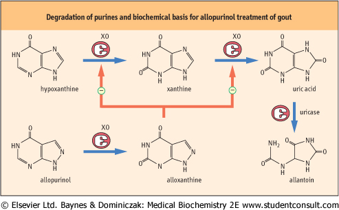
Figure 29.4 Degradation of purines and biochemical basis of allopurine treatment of gout. Inhibition of xanthine oxidase (XO) by alloxanthine is the mechanism involved in allopurinol treatment of gout. The enzyme uricase is missing in primates (including humans), but is commonly used for measurement of serum uric acid levels in humans. treatment of gout. The enzyme uricase is missing in primates (including humans), but is commonly used for measurement of serum uric acid levels in humans. |
| page 417 |  | | page 418 |
| GOUT RESULTS FROM TISSUE ACCUMULATION OF URIC ACID |
| Gout is a disease caused by accumulation of excess uric acid in body fluids (normal range = 0.1-0.5 mmol/L = 2.5-8.0 mg/dL). Uric acid and its urate salts have a low solubility in water, and excessive accumulation of urates results in the precipitation of needle-shaped sodium urate crystals. These crystals are frequently deposited in the soft tissues, particularly in joints. The toxicity of uric acid can be exacerbated by a variety of biochemical defects, including decreased renal clearance, HGPRT deficiency, and glucose-6-phosphatase deficiency. Glucose-6-phosphatase deficiency causes a stimulation of the pentose phosphate pathway, which increases the synthesis of ribose 5-phosphate and consequently PRPP, resulting in overproduction of purines. The over-production of the purines, and their subsequent turnover, leads to an increase in serum uric acid concentration. |
| Gout is often treated with allopurinol, an inhibitor of xanthine oxidase (Fig. 29.4). Xanthine oxidase catalyzes the two-step oxidation of hypoxanthine to uric acid. Allopurinol undergoes the first oxidation to yield alloxanthine, but cannot undergo the second oxidation. Alloxanthine remains bound to the enzyme, thereby inactivating it. This leads to reduced accumulation of uric acid and accumulation of xanthine and hypoxanthine, which are more soluble and are excreted in urine. Increased dietary fiber also appears to suppress serum and urine levels of uric acid by inhibiting the digestion and promoting the excretion of dietary nucleic acids in feces. |
|
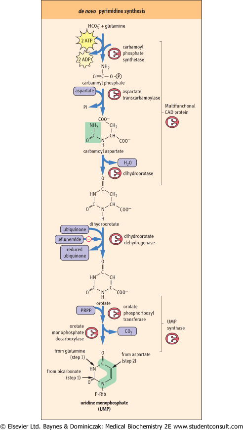
| The pathways of pyrimidine biosynthesis (Fig. 29.5) are invariant in all organisms that have been examined to date. The first step, catalyzed by the enzyme carbamoyl phosphate synthetase, uses bicarbonate, glutamine, and 2 moles of ATP to form carbamoyl phosphate. Carbamoyl phosphate is also used in the synthesis of arginine and in the urea cycle (Chapter 18). Most of the atoms required for formation of the pyrimidine ring are derived from aspartate, added in a single step by aspartate transcarbamoylase (ATCase). Carbamoyl aspartate is then cyclized to dihydroorotic acid by the action of the enzyme dihydroorotase. Dihydroorotic acid is oxidized to orotic acid by a mitochondrial enzyme, dihydroorotate dehydrogenase, which is linked to the electron-transport system through ubiquinone. Leflunomide, a specific inhibitor of this enzyme, is used for treatment of rheumatoid arthritis
because blockage of this step inhibits lymphocyte activation and thereby limits inflammation. The ribosyl-5'-phosphate group from PRPP is transferred onto orotic acid to form orotate monophosphate (OMP). Finally, OMP is decarboxylated to form UMP.
|
| Metabolic channeling by CAD and UMP synthase improves metabolic efficiency
|
| Figure 29.5 The metabolic pathway for the synthesis of pyrimidines. This pathway has two multi-functional proteins. The first, CAD, has three enzymatic activities: Carbamoyl phosphate synthetase, Aspartate transcarbamoylase, and Dihydroorotase (therefore the abbreviation CAD). The second, UMP synthase, has two enzymatic activities: orotate phosphoribosyl transferase and OMP decarboxylase. |
| page 418 | | | page 419 |
| In bacteria and in some fungi, the six enzymes of pyrimidine biosynthesis exist as distinct proteins. However, during the evolution of mammals the first three enzymatic activities have been fused together into a single multifunctional polypeptide. This multifunctional protein, termed CAD for the first letter of each enzyme activity (see legend to Fig. 29.5), is
encoded by a single gene. Similarly, the polypeptides having the final two enzymatic activities of pyrimidine biosynthesis, orotate phosphoribosyl transferase and orotidylate decarboxylase, have also been fused into a single polypeptide, UMP synthase, which is also encoded by a single gene.
|
| The advantage of such multifunctional proteins is that one protein can have multiple sequential enzymatic activities. In such enzymes, the products from the first reaction remain bound to the enzyme and are directed to the second enzymatic center. As with the fatty acid synthase complex, this avoids the diffusion of the metabolic intermediates into the intracellular milieu, thereby improving the metabolic efficiency of the individual steps.
|
|