| Nitrocellulose membranes can store DNA for future use in hybridization studies; Southern blots are the prototype for 'permanent' DNA storage
|
| TISSUE IN SITU HYBRIDIZATION |
A 37-year-old woman was referred for evaluation of excessive hair growth. Endocrine assessment revealed the presence of an adrenal carcinoma secreting cortisol and testosterone . Imaging of the liver revealed the presence of a 2 cm lesion, which was suspect of metastatic spread. Using ultrasound, a biopsy of the liver lesion was obtained, and the lesion studied by in situ hybridization with probes specific to the adrenal cortex. This analysis confirmed that the lesion was a metastasis. . Imaging of the liver revealed the presence of a 2 cm lesion, which was suspect of metastatic spread. Using ultrasound, a biopsy of the liver lesion was obtained, and the lesion studied by in situ hybridization with probes specific to the adrenal cortex. This analysis confirmed that the lesion was a metastasis. |
| Comment. Labeled riboprobes can be used to detect messenger RNA in tissue sections. The probe is often a single-stranded RNA (cRNA) that is complementary to the transcribed messenger RNA (mRNA) of a gene and will thus hybridize with high efficiency to any mRNA present in the tissue section. A cRNA riboprobe can be prepared by in vitro transcription of the gene of interest, which has been cloned into an appropriate plasmid. In order to obtain a complementary RNA to the target, the cloned gene is inserted into the plasmid vector in the reverse orientation relative to a viral promoter. When the gene is transcribed by a viral RNA polymerase, the opposite strand to that normally made in vivo is produced and is therefore complementary to the native mRNA. |
| Hybridization takes place on the microscope slide and the labeled probe will bind to areas of active gene expression. This technique can be also used to study the variation in gene expression in tissues at different times in development, and also tissue-specific gene expression. |
|
Table 34-3.
Types of blots used in molecular biology. |
| Body_ID: None |
| Blots used in molecular biology |
| Body_ID: T034003.50 |
| Blot | Probe | Target |
| Body_ID: T034003.100 |
| Southern | nucleotide | DNA |
| Body_ID: T034003.150 |
| Northern | nucleotide | RNA |
| Body_ID: T034003.200 |
| Western | antibody | protein |
| Body_ID: T034003.250 |
|
| Body_ID: T034003.300 |
Southern and Northern blots use nucleotide-based probes to hybridize to the DNA or RNA on the membrane. Western blots rely on the ability of a specific antibody to bind to a protein of interest.
|
| One of the fundamental steps in the evolution of molecular biology was the discovery that DNA could be transferred from
a semisolid gel onto a nitrocellulose membrane in such a way that the membrane could act as a permanent record of the DNA information in the gel and could be used for multiple-probe experiments. The process whereby the DNA is transferred to the membrane was first described by Ed Southern, but subsequent techniques based on the transfer of RNA and proteins have adopted the direction theme and are called Northern and Western blots, respectively (Table 34.3).
|
| page 477 |  | | page 478 |
| Restriction enzymes are crucial in the process of performing Southern blots
|
| Restriction endonucleases comprise a group of enzymes that cleave double stranded DNA. As discussed later, these enzymes are sequence-specific and each enzyme acts at a limited number of sites in DNA called 'recognition' or 'cutting' sites. These enzymes are an element of the bacterial 'immune system'. Bacteria methylate their own DNA, protecting it from their own restriction enzymes, but cleave infecting viral or bacteriophage DNAs at specific sites, thereby inactivating the virus.
|
|
Table 34-4.
Restriction endonucleases in common use. |
| Body_ID: None |
| Restriction endonucleases |
| Body_ID: T034004.50 |
| Restriction enzyme | Restriction site | Ends |
| Body_ID: T034004.100 |
| HaeIII | GG*CC | flush |
| Body_ID: T034004.150 |
| | CC*GG | |
| Body_ID: T034004.200 |
| MspI | C*CGG | sticky |
| Body_ID: T034004.250 |
| | GGC*C | |
| Body_ID: T034004.300 |
| EcoRV | GAT*ATC | flush |
| Body_ID: T034004.350 |
| | CTA*TAG | |
| Body_ID: T034004.400 |
| EcoRI | G*AATTC | sticky |
| Body_ID: T034004.450 |
| | CTTAA*G | |
| Body_ID: T034004.500 |
| NotI | GC*GGCCGC | sticky |
| Body_ID: T034004.550 |
| | CG CCGG*CG | |
| Body_ID: T034004.600 |
|
| Body_ID: T034004.650 |
Enzymes can cleave DNA to produce 'flush ends' where the DNA is cut 'vertically' leaving two ends that do not have any overhanging nucleotides. However, if the DNA is cleaved 'obliquely', the DNA will have short single stranded overhangs. Such ends are called 'sticky' because they will selectively rejoin (hybridize) to matching overhangs. The sites of cleavage of DNA by restriction enzymes are often described as palindromic because of their inverted repeat symmetry - they have identical sequences in opposite directions on the complementary strands.
|
| RESTRICTION ENZYME CUTTING FREQUENCY |
| Restriction enzymes can cut DNA into fragments at sites determined by the nucleotide sequence of the DNA (Fig. 34.5). The frequency of the cutting sites for various different enzymes varies primarily with the length of the recognition site. Cut sites for an enzymes with a 4-base recognition site, such as HaeIII, would occur by chance once per 256 base pair sequence. Cut sites for an enzyme with an 8-base recognition site, such as NotI, would occur only once in about 65 000 base pairs. As a result of the differing frequencies of cutting sites, there is generally a difference in the size of the cut fragments, frequent cutters generating many small fragments and rare cutters generating fewer and larger fragments. These differences can be exploited when trying to map the location of certain genes to chromosomal locations. |
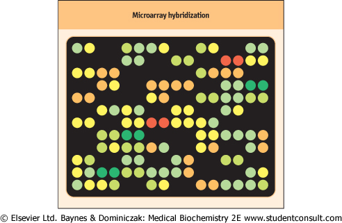
| Figure 34.4 Microarray hybridization. Representation of a small region of an array in which each DNA target dot is duplicated. The probe consisted of a mixture of green-tagged cDNA molecules derived from the mRNA of normal cells and red-tagged molecules derived from the mRNA of diseased cells. The color and brightness of the spots indicates the relative prevalence of particular mRNAs in normal as compared with diseased cells. A yellow signal indicates that a particular mRNA was equally common in both normal and diseased cells. No signal indicates genes whose expression is undetectable. |
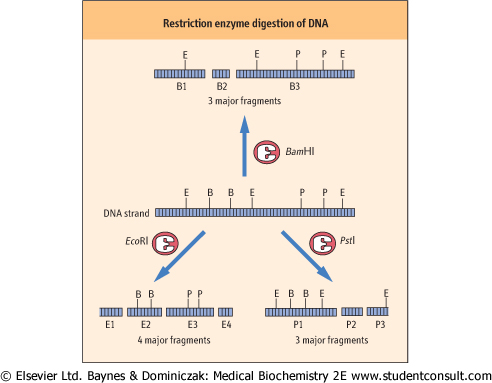
| Figure 34.5 Restriction enzyme digestion of DNA. Digestion of a DNA molecule by several different restriction enzymes may result in many different fragments, even though the apparent size of the fragments is similar. For example, fragments E4 and P2 are of similar size but are clearly different pieces of DNA. E, EcoRI site; B, BamHI site; P, PstI site. |
| page 478 | | | page 479 |
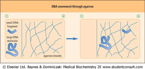
| Figure 34.6 Movement of DNA through agarose during electrophoresis. The molecules must find their way through openings between agarose polymer chains. Small DNA molecules can migrate between the strands with less hindrance. Medium to large DNA molecules take on a random coil geometry and with increasing size have progressively greater difficulty migrating through the polysaccharide network. |
| If DNA is digested by a restriction enzyme, the resulting digested DNA will be reduced to fragments of varying sizes depending on how many cutting sites for that restriction enzyme are present in the DNA. It is important to note that each enzyme will cut DNA into a unique set of fragments (Fig. 34.5).
Originally the location of cut sites and the number of fragments expected were often unknown. However, with the rapid progress in sequencing the genomes of both mammals and other organisms, cut sites can now often be predicted.
|
| Many restriction enzymes recognize sites that are typically four nucleotides (e.g. HaeIII), six nucleotides (e.g. EcoR I) or eight nucleotides (e.g. NotI). Each enzyme recognizes its own site; variation in just one nucleotide within the recognition sequence makes a sequence completely resistant to a particular enzyme.
|
| Blotting techniques involve transfer of DNA, RNA or proteins in the semisolid phase of a gel to a solid phase that may then be used as a template for exposure to a range of molecular probes
|
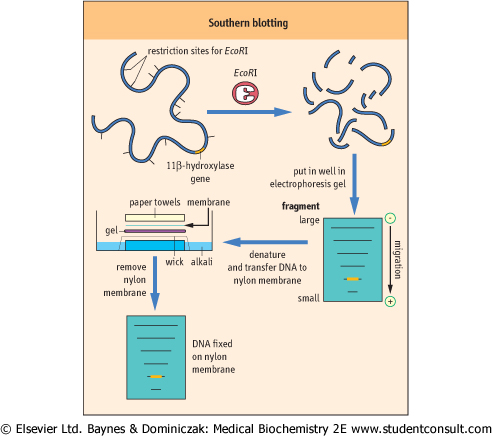
| If DNA is digested by a restriction enzyme, the resulting digest can be separated on the basis of size by gel electrophoresis. A solution of digested DNA is placed in a well in an agarose gel and an electric current applied. The rate of migration of DNA fragments depends on their size, with the smallest fragments moving furthest and the largest moving least (Figure 34.6). Agarose gel electrophoresis is used to separate fragments ranging in size from 100 bases to approximately 20 kb in length (above 40 kb, resolution is minimal). Following electrophoresis, the gels are soaked in a strong alkali solution to render the DNA fragments single-stranded. These single-stranded fragments can then be transferred to a nitrocellulose or nylon membrane to which they bind readily and, if preserved properly, permanently. The process of transfer involves the passage of solute through the gel and into the filter, passively carrying the DNA and producing an image of the gel on the filter (Fig. 34.7).
|
| Figure 34.7 Southern blotting of DNA. DNA digested with a restriction enzyme is size-fractionated by agarose electrophoresis. The agarose gel is then placed in alkali to denature the DNA. The now single-stranded DNA can pass from the gel to the nylon membrane as buffer solution flows upward by capillary action, forming a permanent record of the digested DNA. |
|