| Secondary structure is determined by hydrogen bonding interactions of carbonyl and amide residues in the peptide backbone
|
| The secondary structure of a protein refers to the local structure of the polypeptide chain. This structure is determined by hydrogen bond interactions between the carbonyl oxygen group of one peptide bond and the amide hydrogen of another nearby peptide bond. There are two types of secondary structure, the α-helix and the β-pleated sheet.
|
| page 13 |  | | page 14 |
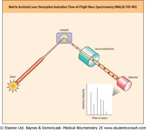
| Figure 2.7 Matrix Assisted Laser Desorption Ionization Time-of-Flight Mass Spectrometry (MALDI-TOF MS). A protein sample is dispersed in a large excess of matrix material that will strongly absorb the incident light. After irradiation by a short laser pulse, the protein is ionised and desorbed from the matrix. The ionized protein molecules are exposed to a voltage gradient in a vacuum and accelerate at a rate that is dependent on their mass to charge ratio. Each molecule reaches the detector after a time (time-of-flight) that can be directly related to its molecular mass. |
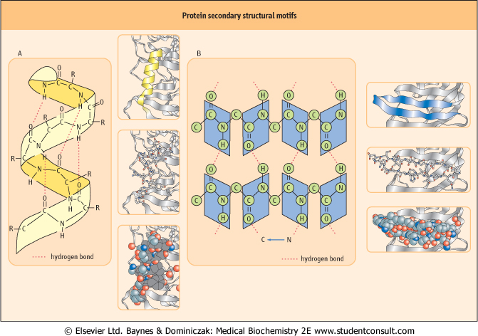
| The α-helix is a rod-like structure with the peptide chain tightly coiled and the side chains of amino acid residues extending outward from the axis of the spiral. Each amide carbonyl group is hydrogen-bonded to the amide hydrogen of a peptide bond that is four residues away along the same chain. There are on average 3.6 amino acid residues per turn of the helix, and the helix winds in a right-handed (clockwise) manner in almost all natural proteins (Fig. 2.8A).
|
| Human genetic defects involving collagen illustrate the close relationship between amino acid sequence and three-dimensional structure. Collagens are the most abundant protein family in the mammalian body, representing about a third of body proteins. Collagens are a major component of connective tissue such as cartilage, tendons, the organic matrix of bones, and the cornea of the eye. |
| Comment. Collagen contains 35% Gly, 11% Ala, and 21% Pro plus Hyp (hydroxyproline). The amino acid sequence in collagen is generally a repeating tripeptide unit, Gly-Xaa-Pro or Gly-Xaa-Hyp, where Xaa can be any amino acid; Hyp =hydroxyproline. This repeating sequence adopts a left-handed helical structure with three residues per turn. Three of these helices wrap around one another with a right-handed twist. The resulting three-stranded molecule is referred to as tropocollagen. Tropocollagen molecules self-assemble into collagen fibrils and are packed together to form collagen fibers. There are metabolic and genetic disorders which result from collagen abnormalities. Scurvy, osteogenesis imperfecta (Chapter 27) and Ehlers-Danlos syndrome result from defects in collagen synthesis and/or crosslinking. |
| page 14 | | | page 15 |
Figure 2.8 Protein secondary structural motifs. (A) An α-helical secondary structure. Hydrogen bonds between 'backbone' amide NH and
 groups stabilize the α-helix. Hydrogen atoms of OH, NH or SH group (hydrogen donors) interact with free electrons of the acceptor atoms such as O, N or S. Even though the bonding energy is lower than that of covalent bonds, they play a pivotal role in the stabilization of protein molecules. R: side chain of amino acids groups stabilize the α-helix. Hydrogen atoms of OH, NH or SH group (hydrogen donors) interact with free electrons of the acceptor atoms such as O, N or S. Even though the bonding energy is lower than that of covalent bonds, they play a pivotal role in the stabilization of protein molecules. R: side chain of amino acids which extend outward from the helix. Ribbon, stick and space-filling models are shown. (B) The parallel β-sheet secondary structure. In the β-conformation, the backbone of the polypeptide chain is extended into a zigzag structure. When the zigzag polypeptide chains are arranged side by side, they form a structure resembling a series of pleats. Ribbon, stick and space-filling models are also shown. which extend outward from the helix. Ribbon, stick and space-filling models are shown. (B) The parallel β-sheet secondary structure. In the β-conformation, the backbone of the polypeptide chain is extended into a zigzag structure. When the zigzag polypeptide chains are arranged side by side, they form a structure resembling a series of pleats. Ribbon, stick and space-filling models are also shown. |
| If the H-bonds are formed between peptide bonds in different chains, the chains become arrayed parallel or antiparallel to one another in what is commonly called a β-pleated sheet. The β-pleated sheet is an extended structure as opposed to the coiled α-helix. It is pleated because the carbon-carbon (C-C) bonds are tetrahedral and cannot exist in a planar configuration. If the polypeptide chain runs in the same direction, it forms a parallel β-sheet (Fig. 2.8B), but in the opposite direction, it forms an antiparallel structure. The β-turn or β-bend refers to the segment in which the polypeptide abruptly reverses direction. Glycine (Gly) and proline
(Pro) residues often occur in β-turns on the surface of globular proteins.
|
|