| Tertiary structure results from folding of the peptide chain
|
| The three-dimensional, folded and biologically active conformation of a protein is referred to as its tertiary structure. This structure reflects the overall shape of the molecule. The tertiary structures of over 1000 proteins have been determined by X-ray crystallography and nuclear magnetic resonance spectroscopy. The folded conformation of proteins that contain more than 200 residues consists of several smaller folded units termed domains.
|
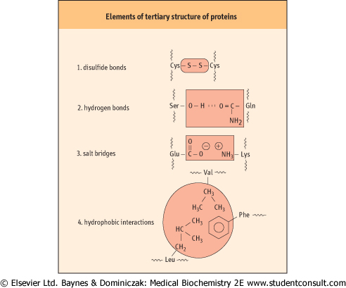
The three-dimensional tertiary structure of a protein is stabilized by interactions between side-chain functional groups, covalent disulfide bonds, hydrogen bonds, salt bridges, and hydrophobic interactions (Fig. 2.8). The side chains of tryptophan and arginine serve as hydrogen donors, whereas asparagine, glutamine, serine, and threonine can serve as both hydrogen donors and acceptors. Lysine, aspartic acid, glutamic acid, tyrosine, and histidine also can serve as both donors and acceptors in the formation of ion pairs (salt bridges). Two opposite-charged amino acids , such as glutamate with a γ-carboxyl group and lysine with an ε-amino group, may form a salt bridge, primarily on the surface of proteins (Fig. 2.9). , such as glutamate with a γ-carboxyl group and lysine with an ε-amino group, may form a salt bridge, primarily on the surface of proteins (Fig. 2.9).
|
| page 15 |  | | page 16 |
| Figure 2.9 Elements of tertiary structure of proteins. Examples of amino acid side-chain interactions contributing to tertiary structure. |
| LENS DISLOCATION IN HOMOCYSTINURIA (INCIDENCE 1 IN 350 000) |
| The most common ocular manifestation of homocystinuria is lens dislocation occurring around age 10 years. |
| Fibrillin, found in the fibres that support the lens, is rich in cysteine residues. Disulphide bonds between these residues are required for the cross-linking and stabilization of protein and lens structure. Homocysteine, a homolog of cysteine, can disrupt these bonds by homocysteine-dependent disulphide exchange. |
| Another equally rare sulfur amino acid disorder - sulfite oxidase deficiency - is also associated with lens dislocation by a similar mechanism (usually presenting at birth with early refractory convulsions). Marfan's syndrome, also associated with lens dislocation, is associated with mutations in the fibrillin gene. |
| Compounds such as urea and guanidine hydrochloride frequently cause denaturation or loss of tertiary structure when
present at high concentrations such as, for example, 8 mol/L urea. These reagents are called denaturants or chaotropic agents.
|
|